

产品中心
当前位置:首页>产品中心Anti-CD5
货号: bs-10218R 基本售价: 1380.0 元 规格: 100ul
- 规格:100ul
- 价格:1380.00元
- 规格:200ul
- 价格:2200.00元
产品信息
- 产品编号
- bs-10218R
- 英文名称
- CD5
- 中文名称
- CD5抗体
- 别 名
- T-cell surface glycoprotein CD5; Lymphocyte antigen 1; Ly-1; Lyt-1; CD-5; CD5 antigen; CD 5; CD5 molecule; CD5 antigen (p56 62); CD5_HUMAN; LEU 1; LEU1; Ly12; LyA; Lymphocyte Antigen CD5; Lymphocyte antigen T1/Leu 1; Lymphocyte antigen T1/Leu-1; Lymphocyte glycoprotein T1/Leu1; OTTHUMP00000236973; p56 62; T1.
- 规格价格
- 100ul/1380元购买 200ul/2200元购买 大包装/询价
- 说 明 书
- 100ul 200ul
- 研究领域
- 免疫学 干细胞 细胞表面分子
- 抗体来源
- Rabbit
- 克隆类型
- Polyclonal
- 交叉反应
- Human, Mouse, Rat, Cow, Horse, Rabbit, Sheep,
- 产品应用
- WB=1:500-2000 ELISA=1:500-1000 IHC-P=1:400-800 IHC-F=1:400-800 Flow-Cyt=1ug/test ICC=1:100-500 IF=1:100-500 (石蜡切片需做抗原修复)
not yet tested in other applications.
optimal dilutions/concentrations should be determined by the end user.
- 分 子 量
- 55kDa
- 细胞定位
- 细胞膜
- 性 状
- Lyophilized or Liquid
- 浓 度
- 1mg/ml
- 免 疫 原
- KLH conjugated synthetic peptide derived from human CD5:221-320/495 <Extracellular>
- 亚 型
- IgG
- 纯化方法
- affinity purified by Protein A
- 储 存 液
- 0.01M TBS(pH7.4) with 1% BSA, 0.03% Proclin300 and 50% Glycerol.
- 保存条件
- Store at -20 °C for one year. Avoid repeated freeze/thaw cycles. The lyophilized antibody is stable at room temperature for at least one month and for greater than a year when kept at -20°C. When reconstituted in sterile pH 7.4 0.01M PBS or diluent of antibody the antibody is stable for at least two weeks at 2-4 °C.
- PubMed
- PubMed
- 产品介绍
- background:
CD5 is a 55kDa T lymphocyte single chain transmembrane glycoprotein. It is present on all mature T lymphocytes, on most thymocytes and on many T cell leukemias and lymphomas. It reacts with a subpopulation of activated B cells. CD5/Lyt1 antigen is a monomeric type I transmembrane glycoprotein expressed on thymocytes, T lymphocytes, and a subset of B lymphocytes, but not on natural killer (NK) cells. It has been identified as the major ligand of the B cell antigen CD72. The frequency of CD5+ B cells exhibits strain dependent variation, and the phenotypic, anatomical, functional, developmental, and pathological characteristics of the CD5+ B cells suggest that they may represent a distinct lineage, known as B1 cells. Binding of CD5 on the T cell surface can augment alloantigen or mitogen induced lymphocyte proliferation and induces increased cytosolic free calcium, IL2 secretion, and IL2R expression. It has been proposed that CD5 negatively regulates signal transduction mediated by the T cell and B cell receptors.
Function:
May act as a receptor in regulating T-cell proliferation.
Subunit:
Interacts with CD72/LYB-2. Interacts with PTPN6/SHP-1.
Subcellular Location:
Cell membrane; Single-pass type I membrane protein.
Post-translational modifications:
Phosphorylated on tyrosine residues by LYN; this creates binding sites for PTPN6/SHP-1.
Similarity:
Contains 3 SRCR domains.
SWISS:
P06127
Gene ID:
921
Database links:Entrez Gene: 921 Human
Omim: 153340 Human
SwissProt: P06127 Human
Unigene: 58685 Human
Important Note:
This product as supplied is intended for research use only, not for use in human, therapeutic or diagnostic applications.
- 产品图片
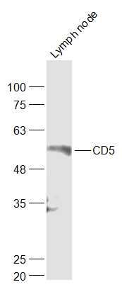 Sample:
Lymph node (Mouse) Lysate at 40 ug
Primary: Anti-CD5 (bs-10218R) at 1/1000 dilution
Secondary: IRDye800CW Goat Anti-Rabbit IgG at 1/20000 dilution
Predicted band size: 55 kD
Observed band size: 55 kD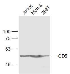 Sample:
Jurkat(Human) Cell Lysate at 30 ug
MOLT-4(Human) Cell Lysate at 30 ug
293T(Human) Cell Lysate at 30 ug
Primary: Anti-CD5 (bs-10218R) at 1/1000 dilution
Secondary: IRDye800CW Goat Anti-Rabbit IgG at 1/20000 dilution
Predicted band size: 55 kD
Observed band size: 55 kD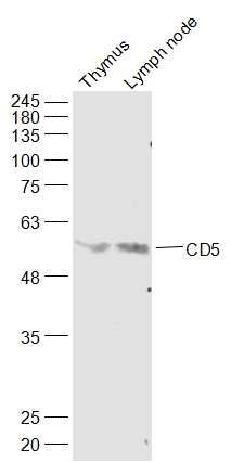 Sample:
Thymus (Mouse) Lysate at 40 ug
Lymph node (Mouse) Lysate at 40 ug
Primary: Anti-CD5 (bs-10218R) at 1/1000 dilution
Secondary: IRDye800CW Goat Anti-Rabbit IgG at 1/20000 dilution
Predicted band size: 55 kD
Observed band size: 55 kD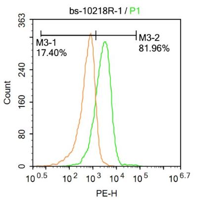 Molt-4 cells were incubated in 5% BSA blocking buffer for 30 min at room temperature. Cells were then stained with CD5 Antibody(bs-10218R)at 1:500 dilution in blocking buffer and incubated for 30 min at room temperature, washed twice with 2%BSA in PBS, followed by secondary antibody incubation for 40 min at room temperature. Acquisitions of 20,000 events were performed. Cells stained with primary antibody (green), and isotype control (orange).