

产品中心
当前位置:首页>产品中心Anti-Phospho-TAK1(Thr184 + Thr187)
货号: bs-3439R 基本售价: 1580.0 元 规格: 100ul
产品信息
- 产品编号
- bs-3439R
- 英文名称
- Phospho-TAK1(Thr184 + Thr187)
- 中文名称
- 磷酸化转化生长因子β活化激酶1
- 别 名
- TAK1(Phospho T184 + T187); TAK1(Phospho Thr184 + Thr187); MAP3K7; Mitogen-activated protein kinase kinase kinase 7; Transforming growth factor-beta-activated kinase 1; TGF-beta-activated kinase 1; MAP3K 7; MAPKKK7; Mitogen activated protein kinase kinase kinase 7; TAK1; TGF beta activated kinase 1; TGF1a; Transforming growth factor beta activated kinase 1; M3K7_HUMAN; Map3k7; MEKK7; TGF-beta-activated kinase 1; TGF1a; Transforming growth factor-beta-activated kinase 1.
- 规格价格
- 100ul/1580元购买 大包装/询价
- 说 明 书
- 100ul
- 产品类型
- 磷酸化抗体
- 研究领域
- 肿瘤 免疫学 信号转导 细胞凋亡 转录调节因子 激酶和磷酸酶
- 抗体来源
- Rabbit
- 克隆类型
- Polyclonal
- 交叉反应
- Human, Mouse, Rat, Chicken, Pig, Cow, Horse, Rabbit,
- 产品应用
- WB=1:1000-2000 ELISA=1:1000-5000 IHC-P=1:50-1000 IHC-F=1:50-1000 IF=1:100-500 (石蜡切片需做抗原修复)
not yet tested in other applications.
optimal dilutions/concentrations should be determined by the end user.
- 分 子 量
- 67kDa
- 细胞定位
- 细胞核 细胞浆 细胞膜
- 性 状
- Lyophilized or Liquid
- 浓 度
- 1mg/ml
- 免 疫 原
- KLH conjugated Synthesised phosphopeptide derived from human TAK1 around the phosphorylation site of Thr184/187:IQ(p-T)HM(p-T)NN
- 亚 型
- IgG
- 纯化方法
- affinity purified by Protein A
- 储 存 液
- 0.01M TBS(pH7.4) with 1% BSA, 0.03% Proclin300 and 50% Glycerol.
- 保存条件
- Store at -20 °C for one year. Avoid repeated freeze/thaw cycles. The lyophilized antibody is stable at room temperature for at least one month and for greater than a year when kept at -20°C. When reconstituted in sterile pH 7.4 0.01M PBS or diluent of antibody the antibody is stable for at least two weeks at 2-4 °C.
- PubMed
- PubMed
- 产品介绍
- background:
The protein encoded by this gene is a member of the serine/threonine protein kinase family. This kinase mediates the signaling transduction induced by TGF beta and morphogenetic protein (BMP), and controls a variety of cell functions including transcription regulation and apoptosis. In response to IL-1, this protein forms a kinase complex including TRAF6, MAP3K7P1/TAB1 and MAP3K7P2/TAB2; this complex is required for the activation of nuclear factor kappa B. This kinase can also activate MAPK8/JNK, MAP2K4/MKK4, and thus plays a role in the cell response to environmental stresses. Four alternatively spliced transcript variants encoding distinct isoforms have been reported. [provided by RefSeq, Jul 2008]
Function:
Component of a protein kinase signal transduction cascade. Mediator of TRAF6 and TGF-beta signal transduction. Activates IKBKB and MAPK8 in response to TRAF6 signaling. Stimulates NF-kappa-B activation and the p38 MAPK pathway. In osmotic stress signaling, plays a major role in the activation of MAPK8/JNK, but not that of NF-kappa-B.
Subunit:
Binds both upstream activators and downstream substrates in multimolecular complexes. Interacts with TAB1/MAP3K7IP1 and TAB2/MAP3K7IP2. Identified in the TRIKA2 complex composed of MAP3K7, TAB1/MAP3K7IP1 and TAB2/MAP3K7IP2. Interacts with PPM1L. Interaction with PP2A and PPP6C leads to its repressed activity. Interacts with TRAF6 and TAB1/MAP3K7IP1; during IL-1 signaling. Interacts with TAOK1 and TAOK2; interaction with TAOK2 interferEs with MAP3K7 interaction with IKKA, thus preventing NF-kappa-B activation. Interacts with WDR34 (via WD domains). Interacts with RBCK1. Interacts with CYLD.
Subcellular Location:
Cytoplasm. Cell membrane; Peripheral membrane protein; Cytoplasmic side. Note=Although the majority of MAP3K7/TAK1 is found in the cytosol, when complexed with TAB1/MAP3K7IP1 and TAB2/MAP3K7IP2, it is also localized at the cell membrane.
Tissue Specificity:
Isoform 1A is the most abundant in ovary, skeletal muscle, spleen and blood mononuclear cells. Isoform 1B is highly expressed in brain, kidney and small intestine. Isoform 1C is the major form in prostate. Isoform 1D is the less abundant form.
Post-translational modifications:
Association with TAB1/MAP3K7IP1 promotes autophosphorylation and subsequent activation. Association with TAB2/MAP3K7IP2, itself associated with free unanchored Lys-63 polyubiquitin chain, promotes autophosphorylation and subsequent activation of MAP3K7. Dephosphorylation at Thr-187 by PP2A and PPP6C leads to inactivation.
Ubiquitinated, leading to proteasomal degradation. Requires Lys-63-linked polyubiquitination for autophosphorylation and subsequent activation. Lys-63-linked ubiquitination does not lead to proteasomal degradation. Deubiquitinated by CYLD, a protease that selectively cleaves Lys-63-linked ubiquitin chains.
Similarity:
Belongs to the protein kinase superfamily. STE Ser/Thr protein kinase family. MAP kinase kinase kinase subfamily.
Contains 1 protein kinase domain.
SWISS:
O43318
Gene ID:
6885
Database links:Entrez Gene: 6885Human
Entrez Gene: 26409Mouse
Entrez Gene: 313121Rat
Omim: 602614Human
SwissProt: O43318Human
SwissProt: Q62073Mouse
SwissProt: P0C8E4Rat
Unigene: 722892Human
Unigene: 258589Mouse
Unigene: 24019Rat
Important Note:
This product as supplied is intended for research use only, not for use in human, therapeutic or diagnostic applications.
- 产品图片
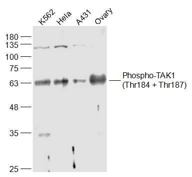 Sample:
K562(Human) Cell Lysate at 30 ug
Hela(Human) Cell Lysate at 30 ug
A431(Human) Cell Lysate at 30 ug
Ovary (Mouse) Lysate at 40 ug
Primary: Anti-Phospho-TAK1(Thr184 + Thr187) (bs-3439R) at 1/1000 dilution
Secondary: IRDye800CW Goat Anti-Rabbit IgG at 1/20000 dilution
Predicted band size: 67 kD
Observed band size: 65 kD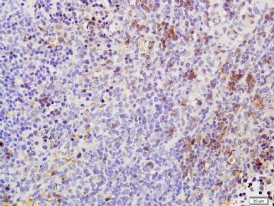 Tissue/cell: rat spleen tissue; 4% Paraformaldehyde-fixed and paraffin-embedded;
Antigen retrieval: citrate buffer ( 0.01M, pH 6.0 ), Boiling bathing for 15min; Block endogenous peroxidase by 3% Hydrogen peroxide for 30min; Blocking buffer (normal goat serum,C-0005) at 37℃ for 20 min;
Incubation: Anti-Phospho-TAK1(Thr184/187) Polyclonal Antibody, Unconjugated(bs-3439R) 1:200, overnight at 4°C, followed by conjugation to the secondary antibody(SP-0023) and DAB(C-0010) staining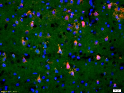 Tissue/cell: rat brain tissue;4% Paraformaldehyde-fixed and paraffin-embedded;
Antigen retrieval: citrate buffer ( 0.01M, pH 6.0 ), Boiling bathing for 15min; Blocking buffer (normal goat serum,C-0005) at 37℃ for 20 min;
Incubation: Anti-Phospho-TAK1(Thr184/187) Polyclonal Antibody, Unconjugated(bs-3439R) 1:200, overnight at 4°C; The secondary antibody was Goat Anti-Rabbit IgG, Cy3 conjugated (bs-0295G-Cy3)used at 1:200 dilution for 40 minutes at 37°C. DAPI(5ug/ml,blue,C-0033) was used to stain the cell nuclei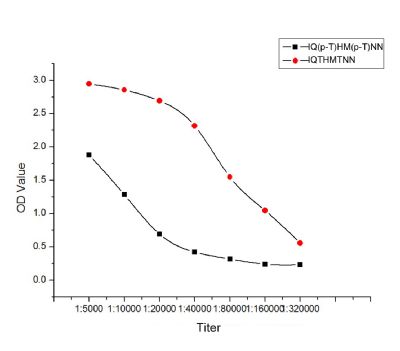 Antigen:
1. IQ(p-T)HM(p-T)NN(red), 0.2ug/100ul
2. IQTHMTNN(black), 0.2ug/100ul
Primary: Antiserum, 1:5000, 1:10000, 1:20000, 1:40000, 1:80000, 1:160000, 1:320000;
Secondary: HRP conjugated Goat Anti-Rabbit IgG(bs-0295G-HRP) at 1: 5000;
TMB(C-0024) staining;
Read the data in MicroplateReader by 450nm.