产品中心
当前位置:首页>产品中心Anti-PSAP
货号: bs-1879R 基本售价: 1380.0 元 规格: 100ul
- 规格:100ul
- 价格:1380.00元
- 规格:200ul
- 价格:2200.00元
产品信息
- 产品编号
- bs-1879R
- 英文名称
- PSAP
- 中文名称
- 鞘脂激活蛋白原抗体
- 别 名
- Prosaposin; A1 activator; Cerebroside sulfate activator; Co-beta-glucosidase; Component C; CSAct; Dispersin; GLBA; Glucosylceramidase activator; Proactivator polypeptide; Proactivator polypeptide precursor; Prosaposin (sphingolipid activator protein 1); prosaposin (variant Gaucher disease and variant metachromatic leukodystrophy); Protein A; Protein C; PSAP; SAP-1; SAP-2; SAP_HUMAN; SAP1; Saposin A; Saposin B; Saposin B Val; Saposin C; Saposin D; Saposin-D; Saposins; Sgp1; Sphingolipid activator protein 1; Sphingolipid activator protein 2; Sulfated glycoprotein 1; Sulfatide/GM1 activator.
- 规格价格
- 100ul/1380元购买 200ul/2200元购买 大包装/询价
- 说 明 书
- 100ul 200ul
- 研究领域
- 肿瘤 发育生物学 神经生物学 细胞周期蛋白 激酶和磷酸酶 脂蛋白 新陈代谢
- 抗体来源
- Rabbit
- 克隆类型
- Polyclonal
- 交叉反应
- Human, Mouse, Rat, Dog,
- 产品应用
- WB=1:500-2000 ELISA=1:500-1000 IHC-P=1:400-800 IHC-F=1:400-800 IF=1:100-500 (石蜡切片需做抗原修复)
not yet tested in other applications.
optimal dilutions/concentrations should be determined by the end user.
- 分 子 量
- 8.8/58kDa
- 细胞定位
- 细胞浆
- 性 状
- Lyophilized or Liquid
- 浓 度
- 1mg/ml
- 免 疫 原
- KLH conjugated synthetic peptide derived from human Prosaposin:101-200/524
- 亚 型
- IgG
- 纯化方法
- affinity purified by Protein A
- 储 存 液
- 0.01M TBS(pH7.4) with 1% BSA, 0.03% Proclin300 and 50% Glycerol.
- 保存条件
- Store at -20 °C for one year. Avoid repeated freeze/thaw cycles. The lyophilized antibody is stable at room temperature for at least one month and for greater than a year when kept at -20°C. When reconstituted in sterile pH 7.4 0.01M PBS or diluent of antibody the antibody is stable for at least two weeks at 2-4 °C.
- PubMed
- PubMed
- 产品介绍
- background:
This gene encodes a highly conserved glycoprotein which is a precursor for 4 cleavage products: saposins A, B, C, and D. Each domain of the precursor protein is approximately 80 amino acid residues long with nearly identical placement of cysteine residues and glycosylation sites. Saposins A-D localize primarily to the lysosomal compartment where they facilitate the catabolism of glycosphingolipids with short oligosaccharide groups. The precursor protein exists both as a secretory protein and as an integral membrane protein and has neurotrophic activities. Mutations in this gene have been associated with Gaucher disease, Tay-Sachs disease, and metachromatic leukodystrophy. Alternative splicing results in multiple transcript variants encoding different isoforms. [provided by RefSeq, Jul 2008]
Function:
The lysosomal degradation of sphingolipids takes place by the sequential action of specific hydrolases. Some of these enzymes require specific low-molecular mass, non-enzymic proteins: the sphingolipids activator proteins (coproteins).
Saposin-A and saposin-C stimulate the hydrolysis of glucosylceramide by beta-glucosylceramidase (EC 3.2.1.45) and galactosylceramide by beta-galactosylceramidase (EC 3.2.1.46). Saposin-C apparently acts by combining with the enzyme and acidic lipid to form an activated complex, rather than by solubilizing the substrate.
Saposin-B stimulates the hydrolysis of galacto-cerebroside sulfate by arylsulfatase A (EC 3.1.6.8), GM1 gangliosides by beta-galactosidase (EC 3.2.1.23) and globotriaosylceramide by alpha-galactosidase A (EC 3.2.1.22). Saposin-B forms a solubilizing complex with the substrates of the sphingolipid hydrolases.
Saposin-D is a specific sphingomyelin phosphodiesterase activator (EC 3.1.4.12).
Subunit:
Saposin-B is a homodimer.
Subcellular Location:
Lysosome.
Post-translational modifications:
This precursor is proteolytically processed to 4 small peptides, which are similar to each other and are sphingolipid hydrolase activator proteins.
N-linked glycans show a high degree of microheterogeneity.
The one residue extended Saposin-B-Val is only found in 5% of the chains.
DISEASE:
Defects in PSAP are the cause of combined saposin deficiency (CSAPD) [MIM:611721]; also known as prosaposin deficiency. CSAPD is due to absence of all saposins, leading to a fatal storage disorder with hepatosplenomegaly and severe neurological involvement.
Defects in PSAP saposin-B region are the cause of leukodystrophy metachromatic due to saposin-B deficiency (MLD-SAPB) [MIM:249900]. MLD-SAPB is an atypical form of metachromatic leukodystrophy. It is characterized by tissue accumulation of cerebroside-3-sulfate, demyelination, periventricular white matter abnormalities, peripheral neuropathy. Additional neurological features include dysarthria, ataxic gait, psychomotr regression, seizures, cognitive decline and spastic quadriparesis.
Defects in PSAP saposin-C region are the cause of atypical Gaucher disease (AGD) [MIM:610539]. Affected individuals have marked glucosylceramide accumulation in the spleen without having a deficiency of glucosylceramide-beta glucosidase characteristic of classic Gaucher disease, a lysosomal storage disorder.
Defects in PSAP saposin-A region are the cause of atypical Krabbe disease (AKRD) [MIM:611722]. AKRD is a disorder of galactosylceramide metabolism. AKRD features include progressive encephalopathy and abnormal myelination in the cerebral white matter resembling Krabbe disease.
Note=Defects in PSAP saposin-D region are found in a variant of Tay-Sachs disease (GM2-gangliosidosis).
Similarity:
Contains 2 saposin A-type domains.
Contains 4 saposin B-type domains.
SWISS:
P07602
Gene ID:
5660
Database links:Entrez Gene: 5660 Human
Entrez Gene: 19156 Mouse
Entrez Gene: 25524 Rat
Omim: 176801 Human
SwissProt: P07602 Human
SwissProt: Q61207 Mouse
SwissProt: P10960 Rat
Unigene: 523004 Human
Unigene: 277498 Mouse
Unigene: 97173 Rat
Important Note:
This product as supplied is intended for research use only, not for use in human, therapeutic or diagnostic applications.
表达在正常的前列腺、前列腺增生和前列腺肿瘤中。
- 产品图片
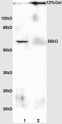
 Protein:
testis(rat) lysates at 30ug;
brain(rat) lysates at 30ug;
Primary: Anti-Anti-PSAP/PAP (bs-1879R) at 1:200;
Secondary: HRP conjugated Goat Anti-Rabbit IgG(bs-0295G-HRP) at 1: 3000;
ECL excitated the fluorescence;
Predicted band size : 58kD
Observed band size : 58kD Sample:
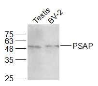
Testis (Rat) Lysate at 40 ug
BV-2 (Mouse) CellLysate at 30 ug
Primary: Anti-PSAP (bs-1879R) at 1/1000 dilution
Secondary: IRDye800CW Goat Anti-Rabbit IgG at 1/20000 dilution
Predicted band size: 8.8/58 kD
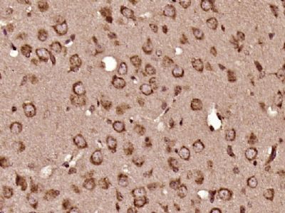
Observed band size: 58 kD Paraformaldehyde-fixed, paraffin embedded (Mouse brain); Antigen retrieval by boiling in sodium citrate buffer (pH6.0) for 15min; Block endogenous peroxidase by 3% hydrogen peroxide for 20 minutes; Blocking buffer (normal goat serum) at 37°C for 30min; Antibody incubation with (PSAP) Polyclonal Antibody, Unconjugated (bs-1879R) at 1:400 overnight at 4°C, followed by operating according to SP Kit(Rabbit) (sp-0023) instructionsand DAB staining.
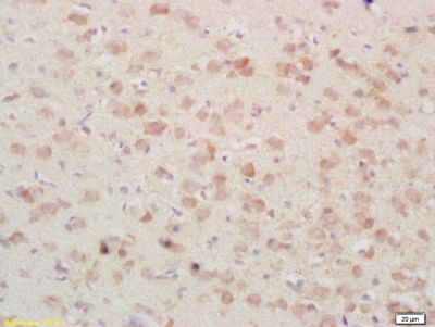
 Tissue/cell: rat brain tissue; 4% Paraformaldehyde-fixed and paraffin-embedded;
Antigen retrieval: citrate buffer ( 0.01M, pH 6.0 ), Boiling bathing for 15min; Block endogenous peroxidase by 3% Hydrogen peroxide for 30min; Blocking buffer (normal goat serum,C-0005) at 37℃ for 20 min;
Incubation: Anti-PSAP/PAP Polyclonal Antibody, Unconjugated(bs-1879R) 1:200, overnight at 4°C, followed by conjugation to the secondary antibody(SP-0023) and DAB(C-0010) staining

