

产品中心
当前位置:首页>产品中心Anti-VE Cadherin
货号: bs-4310R 基本售价: 780.0 元 规格: 50ul
- 规格:50ul
- 价格:780.00元
- 规格:100ul
- 价格:1380.00元
- 规格:200ul
- 价格:2200.00元
产品信息
- 产品编号
- bs-4310R
- 英文名称
- VE Cadherin
- 中文名称
- 血管内皮钙粘蛋白抗体
- 别 名
- VE-cadherin; cadherin-5; VE-Cadherin;Cdh5; Vascular endothelial cadherin; 7B4 antigen; CD144 antigen; VECD; Vascular endothelial cell cadherin; 7B 4; 7B4; 7B4 antigen; Cadherin 5; Cadherin 5 type 2; Cadherin-5; Cadherin5; CD 144; CD144; CD144 antigen; CDH 5; CDH5; CDH5 protein; Vascular endothelial cadherin; Vascular epithelium cadherin; VE Cad antibody VE-cadherin; VE cadherin; VEC; CADH5_HUMAN.
- 规格价格
- 50ul/780元购买 100ul/1380元购买 200ul/2200元购买 大包装/询价
- 说 明 书
- 50ul 100ul 200ul
- 研究领域
- 肿瘤 心血管 细胞生物 信号转导 细胞粘附分子 血管内皮细胞 内皮细胞 细胞骨架 细胞外基质
- 抗体来源
- Rabbit
- 克隆类型
- Polyclonal
- 交叉反应
- Human, Mouse, Rat, Pig, Cow, Horse,
- 产品应用
- Flow-Cyt=1μg/Test (石蜡切片需做抗原修复)
not yet tested in other applications.
optimal dilutions/concentrations should be determined by the end user.
- 分 子 量
- 81kDa
- 细胞定位
- 细胞膜
- 性 状
- Lyophilized or Liquid
- 浓 度
- 1mg/ml
- 免 疫 原
- KLH conjugated synthetic peptide derived from human VE Cadherin:251-320/784 <Extracellular>
- 亚 型
- IgG
- 纯化方法
- affinity purified by Protein A
- 储 存 液
- 0.01M TBS(pH7.4) with 1% BSA, 0.03% Proclin300 and 50% Glycerol.
- 保存条件
- Store at -20 °C for one year. Avoid repeated freeze/thaw cycles. The lyophilized antibody is stable at room temperature for at least one month and for greater than a year when kept at -20°C. When reconstituted in sterile pH 7.4 0.01M PBS or diluent of antibody the antibody is stable for at least two weeks at 2-4 °C.
- PubMed
- PubMed
- 产品介绍
- background:
This gene is a classical cadherin from the cadherin superfamily and is located in a six-cadherin cluster in a region on the long arm of chromosome 16 that is involved in loss of heterozygosity events in breast and prostate cancer. The encoded protein is a calcium-dependent cell-cell adhesion glycoprotein comprised of five extracellular cadherin repeats, a transmembrane region and a highly conserved cytoplasmic tail. Functioning as a classic cadherin by imparting to cells the ability to adhere in a homophilic manner, the protein may play an important role in endothelial cell biology through control of the cohesion and organization of the intercellular junctions. An alternative splice variant has been described but its full length sequence has not been determined. [provided by RefSeq, Jul 2008].
Function:
Cadherins are calcium dependent cell adhesion proteins. They preferentially interact with themselves in a homophilic manner in connecting cells; cadherins may thus contribute to the sorting of heterogeneous cell types. This cadherin may play a important role in endothelial cell biology through control of the cohesion and organization of the intercellular junctions. It associates with alpha-catenin forming a link to the cytoskeleton.
Subunit:
Interacts via cadherin 5 domain with PTPRB. Interacts with TRPC4. Interacts with KRIT1.
Subcellular Location:
Cell junction. Cell membrane. Found at cell-cell boundaries and probably at cell-matrix boundaries.
Tissue Specificity:
Endothelial tissues and brain.
Post-translational modifications:
Phosphorylated on tyrosine residues by KDR/VEGFR-2. Dephosphorylated by PTPRB.
Similarity:
Contains 5 cadherin domains.
SWISS:
P33151
Gene ID:
1003
Database links:Entrez Gene: 1003 Human
Entrez Gene: 12562 Mouse
Entrez Gene: 307618 Rat
Omim: 601120 Human
SwissProt: P33151 Human
SwissProt: P55284 Mouse
Unigene: 76206 Human
Unigene: 21767 Mouse
Unigene: 224644 Rat
Important Note:
This product as supplied is intended for research use only, not for use in human, therapeutic or diagnostic applications.
- 产品图片
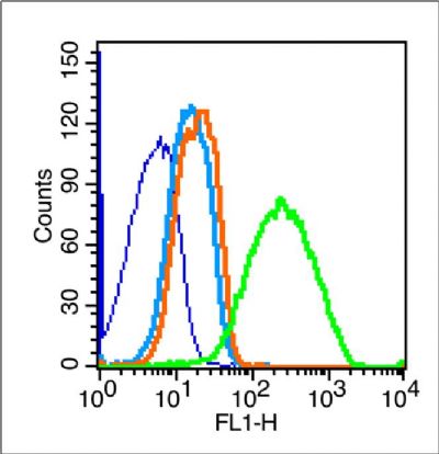 Blank control (blue line): Mouse kidney (blue).
Primary Antibody (green line): Rabbit Anti-VE Cadherin antibody (bs-4310R)
Dilution: 1μg /10^6 cells;
Isotype Control Antibody (orange line): Rabbit IgG .
Secondary Antibody (white blue line): F(ab’)2 fragment goat anti-rabbit IgG-FITC
Dilution: 1μg /test.
Protocol
The cells were fixed with 70% ice-cold methanol overnight at 4℃.Cells stained with Primary Antibody for 30 min at room temperature. The cells were then incubated in 1 X PBS/2%BSA/10% goat serum to block non-specific protein-protein interactions followed by the antibody for 15 min at room temperature. The secondary antibody used for 40 min at room temperature. Acquisition of 20,000 events was performed.