

产品中心
当前位置:首页>产品中心Anti-PKC beta 1 + 2
货号: bs-0267R 基本售价: 380.0 元 规格: 20ul
- 规格:20ul
- 价格:380.00元
- 规格:50ul
- 价格:780.00元
- 规格:100ul
- 价格:1380.00元
- 规格:200ul
- 价格:2200.00元
产品信息
- 产品编号
- bs-0267R
- 英文名称
- PKC beta 1 + 2
- 中文名称
- 蛋白激酶C beta 1/2抗体
- 别 名
- PKC beta 1 + PKC beta 2; PKCB; PKC B; PKCB1; PKCB2; PRKCB; PRKCB I; PRKCB II; PRKCB1; PRKCB2; Protein kinase C beta 1; Protein kinase C beta 2; Protein kinase C beta; PKC beta 1; PKC beta 2; KPCB_HUMAN; MGC41878; PKC beta; PKC-B; PKC-beta; PRKC B; PRKC B1; Protein kinase C beta 1; Protein kinase C beta; Protein kinase C beta type.
- 规格价格
- 50ul/780元购买 100ul/1380元购买 200ul/2200元购买 大包装/询价
- 说 明 书
- 50ul 100ul 200ul
- 研究领域
- 细胞生物 信号转导 激酶和磷酸酶
- 抗体来源
- Rabbit
- 克隆类型
- Polyclonal
- 交叉反应
- Human, Mouse, Rat, Pig, Cow, Rabbit,
- 产品应用
- WB=1:500-2000 ELISA=1:500-1000 IHC-P=1:400-800 IHC-F=1:400-800 Flow-Cyt=1:ug/Test IF=1:100-500 (石蜡切片需做抗原修复)
not yet tested in other applications.
optimal dilutions/concentrations should be determined by the end user.
- 分 子 量
- 74kDa
- 细胞定位
- 细胞核 细胞浆 细胞膜
- 性 状
- Lyophilized or Liquid
- 浓 度
- 1mg/ml
- 免 疫 原
- KLH conjugated synthetic peptide derived from human PKC beta 1:601-673/673
- 亚 型
- IgG
- 纯化方法
- affinity purified by Protein A
- 储 存 液
- 0.01M TBS(pH7.4) with 1% BSA, 0.03% Proclin300 and 50% Glycerol.
- 保存条件
- Store at -20 °C for one year. Avoid repeated freeze/thaw cycles. The lyophilized antibody is stable at room temperature for at least one month and for greater than a year when kept at -20°C. When reconstituted in sterile pH 7.4 0.01M PBS or diluent of antibody the antibody is stable for at least two weeks at 2-4 °C.
- PubMed
- PubMed
- 产品介绍
- background:
Protein kinase C (PKC) is a family of serine- and threonine-specific protein kinases that can be activated by calcium and the second messenger diacylglycerol. PKC family members phosphorylate a wide variety of protein targets and are known to be involved in diverse cellular signaling pathways. PKC family members also serve as major receptors for phorbol esters, a class of tumor promoters. Each member of the PKC family has a specific expression profile and is believed to play a distinct role. The protein encoded by this gene is one of the PKC family members. It is a calcium-independent and phospholipid-dependent protein kinase. This kinase is important for T-cell activation. It is required for the activation of the transcription factors NF-kappaB and AP-1, and may link the T cell receptor (TCR) signaling complex to the activation of the transcription factors.
Function:
Calcium-activated and phospholipid-dependent serine/threonine-protein kinase involved in various processes such as regulation of the B-cell receptor (BCR) signalosome, apoptosis and transcription regulation. Plays a key role in B-cell activation and function by regulating BCR-induced NF-kappa-B activation and B-cell suvival. Required for recruitment and activation of the IKK kinase to lipid rafts and mediates phosphorylation of CARD11/CARMA1 at Ser-559, Ser-644 and Ser-652, leading to activate the NF-kappa-B signaling. Involved in apoptosis following oxidative damage: in case of oxidative conditions, specifically phosphorylates Ser-36 of isoform p66Shc of SHC1, leading to mitochondrial accumulation of p66Shc, where p66Shc acts as a reactive oxygen species producer. Acts as a coactivator of androgen receptor (ANDR)-dependent transcription, by being recruited to ANDR target genes and specifically mediating phosphorylation of Thr-6 of histone H3 (H3T6ph), a specific tag for epigenetic transcriptional activation that prevents demethylation of histone H3 Lys-4 (H3K4me) by LSD1/KDM1A. Also involved in triglyceride homeostasis. Serves as the receptor for phorbol esters, a class of tumor promoters.
Subunit:
Recruited in a circadian manner into a nuclear complex which also includes BMAL1 and GNB2L1/RACK1 (By similarity). Interacts with ADAP1/CENTA1, CSPG4 and PRKCABP. Binds to SDPR in the presence of phosphatidylserine. Interacts with PICK1 (via PDZ domain). Interacts with TRIM41.
Subcellular Location:
Cytoplasm. Cell membrane; Peripheral membrane protein. Mitochondrion membrane; Peripheral membrane protein (Probable). Nucleus.
Post-translational modifications:
Phosphorylation on Thr-500 within the activation loop renders it competent to autophosphorylate. Subsequent autophosphorylation of Thr-642 maintains catalytic competence, and autophosphorylation on Ser-661 appears to release the kinase into the cytosol. Autophosphorylation on other sites i.e. in the N-terminal and hinge regions have no effect on enzyme activity.
Similarity:
Belongs to the protein kinase superfamily. AGC Ser/Thr protein kinase family. PKC subfamily.
Contains 1 AGC-kinase C-terminal domain.
Contains 1 C2 domain.
Contains 2 phorbol-ester/DAG-type zinc fingers.
Contains 1 protein kinase domain.
SWISS:
P05771
Gene ID:
5579
Database links:Entrez Gene: 5579Human
Entrez Gene: 18751Mouse
Entrez Gene: 25023Rat
Omim: 176970Human
SwissProt: P05771Human
SwissProt: P68404Mouse
SwissProt: P68403Rat
Unigene: 460355Human
Unigene: 207496Mouse
Unigene: 446371Mouse
Unigene: 91118Rat
Important Note:
This product as supplied is intended for research use only, not for use in human, therapeutic or diagnostic applications.
- 产品图片
 Sample:
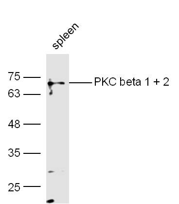
spleen (Mouse) Lysate at 40 ug
Primary: Anti-PKC beta 1 + 2 (bs-0267R) at 1/300 dilution
Secondary: IRDye800CW Goat Anti-Rabbit IgG at 1/20000 dilution
Predicted band size: 74 kD
Observed band size: 74 kD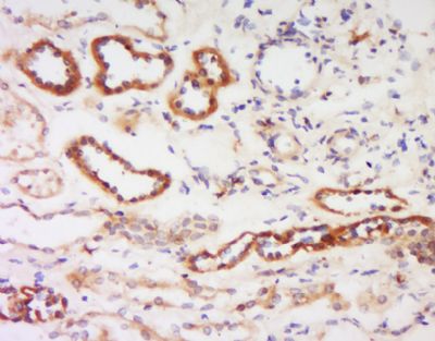 Tissue/cell: human kidney tissue; 4% Paraformaldehyde-fixed and paraffin-embedded;
Antigen retrieval: citrate buffer ( 0.01M, pH 6.0 ), Boiling bathing for 15min; Block endogenous peroxidase by 3% Hydrogen peroxide for 30min; Blocking buffer (normal goat serum,C-0005) at 37℃ for 20 min;
Incubation: Anti- PKC beta-1+2 Polyclonal Antibody, Unconjugated(bs-0267R) 1:200, overnight at 4°C, followed by conjugation to the secondary antibody(SP-0023) and DAB(C-0010) staining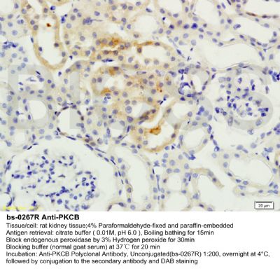
 Blank control (Black line): Molt4 (Black).
Primary Antibody (green line):Rabbit Anti-PKC beta 1 + 2 antibody (bs-0267R)
Dilution:1μg /10^6 cells;
Isotype Control Antibody (orange line): Rabbit IgG .
Secondary Antibody (white blue line): Goat anti-rabbit IgG-AF647
Dilution: 1μg /test.
Protocol
The cells were fixed with 4% PFA (10min at room temperature)and then permeabilized with 90% ice-cold methanol for 20 min at room temperature. The cells were then incubated in 5%BSA to block non-specific protein-protein interactions for 30 min at room temperature .Cells stained with Primary Antibody for 30 min at room temperature. The secondary antibody used for 40 min at room temperature. Acquisition of 20,000 events was performed.