产品中心
当前位置:首页>产品中心Anti-PDGF Receptor beta
货号: bs-0232R 基本售价: 780.0 元 规格: 50ul
- 规格:50ul
- 价格:780.00元
- 规格:100ul
- 价格:1380.00元
- 规格:200ul
- 价格:2200.00元
产品信息
- 产品编号
- bs-0232R
- 英文名称
- PDGF Receptor beta
- 中文名称
- 血小板源性生长因子受体B/PDGFRβ抗体
- 别 名
- CD140B; Beta platelet derived growth factor receptor; Beta-type platelet-derived growth factor receptor; CD 140B; CD140 antigen-like family member B; CD140B; CD140b antigen; JTK12; OTTHUMP00000160528; PDGF R beta; PDGF-R-beta; PDGFR; PDGFR1; PDGFRB; PGFRB_HUMAN; Platelet derived growth factor receptor beta; Platelet derived growth factor receptor beta polypeptide; PDGF Receptor beta; beta-type platelet-derived growth factor receptor precursor; PDGFRβ; PDGF Rβ; PDGF Receptor β.
- 规格价格
- 50ul/780元购买 100ul/1380元购买 200ul/2200元购买 大包装/询价
- 说 明 书
- 50ul 100ul 200ul
- 研究领域
- 肿瘤 细胞生物 信号转导 细胞凋亡 生长因子和激素 细胞膜受体
- 抗体来源
- Rabbit
- 克隆类型
- Polyclonal
- 交叉反应
- Human, Mouse, Rat, Dog,
- 产品应用
- WB=1:500-2000 ELISA=1:500-1000 IHC-P=1:400-800 IHC-F=1:400-800 ICC=1:100-500 IF=1:100-500 (石蜡切片需做抗原修复)
not yet tested in other applications.
optimal dilutions/concentrations should be determined by the end user.
- 分 子 量
- 118kDa
- 细胞定位
- 细胞膜
- 性 状
- Lyophilized or Liquid
- 浓 度
- 1mg/ml
- 免 疫 原
- KLH conjugated synthetic peptide derived from human PDGF-R-B:981-1066/1106 <Cytoplasmic>
- 亚 型
- IgG
- 纯化方法
- affinity purified by Protein A
- 储 存 液
- 0.01M TBS(pH7.4) with 1% BSA, 0.03% Proclin300 and 50% Glycerol.
- 保存条件
- Store at -20 °C for one year. Avoid repeated freeze/thaw cycles. The lyophilized antibody is stable at room temperature for at least one month and for greater than a year when kept at -20°C. When reconstituted in sterile pH 7.4 0.01M PBS or diluent of antibody the antibody is stable for at least two weeks at 2-4 °C.
- PubMed
- PubMed
- 产品介绍
- background:
The PDGF Receptor Type A (Alpha platelet-derived growth factor receptor precursor, CD140a antigen), a 170kD protein, binds all three isoforms of PDGF with high affinity whereas the PDGF Receptor Type B, a 190kD protein, appears to bind only the PDGF BB homodimer with high affinity. Both receptors are transmembranous, ligand activated protein tyrosine kinases, which phosphorylate a number of important signal transduction proteins, which are bound with differential affinities via SH2 domains. The response of any given cell to PDGF will depend on the types of receptors displayed on the surface and isoforms of PDGF present in the extracellular environment.
Function:
Receptor that binds specifically to PDGFB and PDGFD and has a tyrosine-protein kinase activity. Phosphorylates Tyr residues at the C-terminus of PTPN11 creating a binding site for the SH2 domain of GRB2.
Subunit:
Interacts with homodimeric PDGFB and PDGFD, and with heterodimers formed by PDGFA and PDGFB. May also interact with homodimeric PDGFC. Monomer in the absence of bound ligand. Interaction with homodimeric PDGFB, heterodimers formed by PDGFA and PDGFB or homodimeric PDGFD, leads to receptor dimerization, where both PDGFRA homodimers and heterodimers with PDGFRB are observed. Interacts with SH2B2/APS. Interacts directly (tyrosine phosphorylated) with SHB. Interacts (tyrosine phosphorylated) with PIK3R1. Interacts (tyrosine phosphorylated) with CBL. Interacts (tyrosine phosphorylated) with SRC and SRC family kinases. Interacts (tyrosine phosphorylated) with PIK3C2B, maybe indirectly. Interacts (tyrosine phosphorylated) with SHC1, GRB7, GRB10 and NCK1. Interaction with GRB2 is mediated by SHC1. Interacts (via C-terminus) with SLC9A3R1.
Subcellular Location:
Cell membrane; Single-pass type I membrane protein. Cytoplasmic vesicle. Lysosome lumen. Note=After ligand binding, the autophosphorylated receptor is ubiquitinated and internalized, leading to its degradation.
Post-translational modifications:
Autophosphorylated on tyrosine residues upon ligand binding. Autophosphorylation occurs in trans, i.e. one subunit of the dimeric receptor phosphorylates tyrosine residues on the other subunit. Phosphorylation at Tyr-579, and to a lesser degree, at Tyr-581, is important for interaction with SRC family kinases. Phosphorylation at Tyr-740 and Tyr-751 is important for interaction with PIK3R1. Phosphorylation at Tyr-751 is important for interaction with NCK1. Phosphorylation at Tyr-771 and Tyr-857 is important for interaction with RASA1/GAP. Phosphorylation at Tyr-857 is important for efficient phosphorylation of PLCG1 and PTPN11, resulting in increased phosphorylation of AKT1, MAPK1/ERK2 and/or MAPK3/ERK1, PDCD6IP/ALIX and STAM, and in increased cell proliferation. Phosphorylation at Tyr-1009 is important for interaction with PTPN11. Phosphorylation at Tyr-1009 and Tyr-1021 is important for interaction with PLCG1. Phosphorylation at Tyr-1021 is important for interaction with CBL; PLCG1 and CBL compete for the same binding site. Dephosphorylated by PTPRJ at Tyr-751, Tyr-857, Tyr-1009 and Tyr-1021.
N-glycosylated.
Ubiquitinated. After autophosphorylation, the receptor is polyubiquitinated, leading to its degradation.
DISEASE:
Note=A chromosomal aberration involving PDGFRB is found in a form of chronic myelomonocytic leukemia (CMML). Translocation t(5;12)(q33;p13) with EVT6/TEL. It is characterized by abnormal clonal myeloid proliferation and by progression to acute myelogenous leukemia (AML).
Note=A chromosomal aberration involving PDGFRB may be a cause of acute myelogenous leukemia. Translocation t(5;14)(q33;q32) with TRIP11. The fusion protein may be involved in clonal evolution of leukemia and eosinophilia.
Note=A chromosomal aberration involving PDGFRB may be a cause of juvenile myelomonocytic leukemia. Translocation t(5;17)(q33;p11.2) with SPECC1.
Defects in PDGFRB are a cause of myeloproliferative disorder chronic with eosinophilia (MPE) [MIM:131440]. A hematologic disorder characterized by malignant eosinophils proliferation. Note=A chromosomal aberration involving PDGFRB is found in many instances of myeloproliferative disorder chronic with eosinophilia. Translocation t(5;12) with ETV6 on chromosome 12 creating an PDGFRB-ETV6 fusion protein. Translocation t(5;15)(q33;q22) with TP53BP1 creating a PDGFRB-TP53BP1 fusion protein.
Note=A chromosomal aberration involving PDGFRB may be the cause of a myeloproliferative disorder (MBD) associated with eosinophilia. Translocation t(1;5)(q23;q33) that forms a PDE4DIP-PDGFRB fusion protein.
Note=A chromosomal aberration involving PGFRB is found in a patient with T-lymphoblastic lymphoma (T-ALL) and an associated myeloproliferative neoplasm (MPN) with eosinophilia. Translocation t(5;6)(q33-34;q23) with CEP85L. The translocation fuses the 5-end of CEP85L (isoform 4) to the 3-end of PDGFRB.
Similarity:
Belongs to the protein kinase superfamily. Tyr protein kinase family. CSF-1/PDGF receptor subfamily.
Contains 5 Ig-like C2-type (immunoglobulin-like) domains.
Contains 1 protein kinase domain.
SWISS:
P09619
Gene ID:
5159
Database links:Entrez Gene: 5159Human
Entrez Gene: 18596Mouse
Entrez Gene: 24629Rat
Omim: 173410Human
SwissProt: P09619Human
SwissProt: P05622Mouse
SwissProt: Q05030Rat
Unigene: 509067Human
Unigene: 4146Mouse
Unigene: 98311Rat
Important Note:
This product as supplied is intended for research use only, not for use in human, therapeutic or diagnostic applications.
- 产品图片
 Sample:
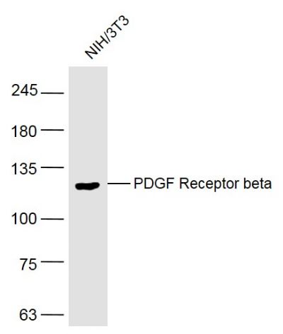
NIH/3T3(Rat) Cell Lysate at 30 ug
Primary: Anti-PDGF Receptor beta (bs-0232R) at 1/300 dilution
Secondary: IRDye800CW Goat Anti-Rabbit IgG at 1/20000 dilution
Predicted band size: 118 kD
Observed band size: 118 kD Sample:
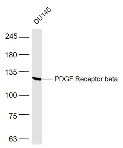
DU145(Human) Cell Lysate at 30 ug
Primary: Anti-PDGF Receptor beta (bs-0232R) at 1/300 dilution
Secondary: IRDye800CW Goat Anti-Rabbit IgG at 1/20000 dilution
Predicted band size: 118 kD
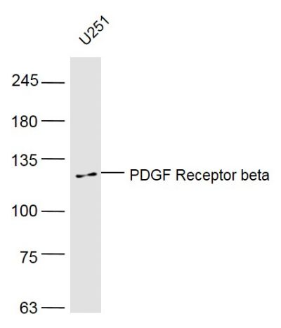
Observed band size: 118 kD Sample:
U251(Human) Cell Lysate at 30 ug
Primary: Anti-PDGF Receptor beta (bs-0232R) at 1/300 dilution
Secondary: IRDye800CW Goat Anti-Rabbit IgG at 1/20000 dilution
Predicted band size: 118 kD
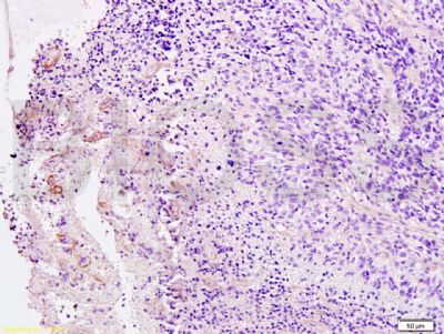
Observed band size: 118 kD Tissue/cell: Human brain glioma; 4% Paraformaldehyde-fixed and paraffin-embedded;
Antigen retrieval: citrate buffer ( 0.01M, pH 6.0 ), Boiling bathing for 15min; Block endogenous peroxidase by 3% Hydrogen peroxide for 30min; Blocking buffer (normal goat serum,C-0005) at 37℃ for 20 min;
Incubation: Anti-PDGFRB Polyclonal Antibody, Unconjugated(bs-0232R) 1:200, overnight at 4°C, followed by conjugation to the secondary antibody(SP-0023) and DAB(C-0010) staining

