产品中心
当前位置:首页>产品中心Anti-MUC1
货号: bs-1018R 基本售价: 1380.0 元 规格: 100ul
- 规格:100ul
- 价格:1380.00元
- 规格:200ul
- 价格:2200.00元
产品信息
- 产品编号
- bs-1018R
- 英文名称
- MUC1
- 中文名称
- 粘蛋白-1抗体
- 别 名
- Breast carcinoma associated antigen DF3; CA 15 3; CA15-3; CA153; CA15-3 antigen; CA15.3; CA-153; Carcinoma associated mucin; CD 227; CD227; CD227 antigen; DF3 antigen; EMA; Episialin; Epithelial membrane antigen; Epithelial mucin tandem repeat sequence; H23 antigen; H23AG; HGNC:7508; MAM6; MUC 1; MUC-1; Mucin 1; Mucin 1 precursor; Mucin1; Mucin 1; Peanut reactive urinary mucin; PEM; PEMT; Polymorphic epithelial mucin; PUM; Tumor associated epithelial membrane antigen; Tumor associated mucin.
- 规格价格
- 100ul/1380元购买 200ul/2200元购买 大包装/询价
- 说 明 书
- 100ul 200ul
- 研究领域
- 肿瘤 免疫学 生长因子和激素 细胞粘附分子
- 抗体来源
- Rabbit
- 克隆类型
- Polyclonal
- 交叉反应
- Human, Rabbit,
- 产品应用
- IHC-P=1:400-800 IHC-F=1:400-800 Flow-Cyt=3μg/Test IF=1:100-500 (石蜡切片需做抗原修复)
not yet tested in other applications.
optimal dilutions/concentrations should be determined by the end user.
- 分 子 量
- 136kDa
- 细胞定位
- 细胞核 细胞浆 细胞膜 分泌型蛋白
- 性 状
- Lyophilized or Liquid
- 浓 度
- 1mg/ml
- 免 疫 原
- KLH conjugated synthetic peptide derived from human Muc1:151-250/1255 <Extracellular>
- 亚 型
- IgG
- 纯化方法
- affinity purified by Protein A
- 储 存 液
- 0.01M TBS(pH7.4) with 1% BSA, 0.03% Proclin300 and 50% Glycerol.
- 保存条件
- Store at -20 °C for one year. Avoid repeated freeze/thaw cycles. The lyophilized antibody is stable at room temperature for at least one month and for greater than a year when kept at -20°C. When reconstituted in sterile pH 7.4 0.01M PBS or diluent of antibody the antibody is stable for at least two weeks at 2-4 °C.
- PubMed
- PubMed
- 产品介绍
- background:
This gene is a member of the mucin family and encodes a membrane bound, glycosylated phosphoprotein. The protein is anchored to the apical surface of many epithelia by a transmembrane domain, with the degree of glycosylation varying with cell type. It
also includes a 20 aa variable number tandem repeat (VNTR) domain, with the number of repeats varying from 20 to 120 in different individuals. The protein serves a protective function by binding to pathogens and also functions in a cell signaling capacity. Overexpression, aberrant intracellular localization, and changes in glycosylation of this protein have been associated with carcinomas. Multiple alternatively spliced transcript variants that encode different isoforms of this gene have been reported, but the full-length nature of only some has been determined.
Function:
The alpha subunit has cell adhesive properties. Can act both as an adhesion and an anti-adhesion protein. May provide a protective layer on epithelial cells against bacterial and enzyme attack.
The beta subunit contains a C-terminal domain which is involved in cell signaling, through phosphorylations and protein-protein interactions. Modulates signaling in ERK, SRC and NF-kappa-B pathways. In activated T-cells, influences directly or indirectly the Ras/MAPK pathway. Promotes tumor progression. Regulates TP53-mediated transcription and determines cell fate in the genotoxic stress response. Binds, together with KLF4, the PE21 promoter element of TP53 and represses TP53 activity.
Subunit:
The alpha subunit forms a tight, non-covalent heterodimeric complex with the proteolytically-released beta-subunit. Interaction, via the tandem repeat region, with domain 1 of ICAM1 is implicated in cell migration and metastases. Isoform 1 binds directly the SH2 domain of GRB2, and forms a MUC1/GRB2/SOS1 complex involved in RAS signaling. The cytoplasmic tail (MUC1CT) interacts with several proteins such as SRC, CTNNB1 and ERBs. Interaction with the SH2 domain of CSK decreases interaction with GSK3B. Interacts with CTNNB1/beta-catenin and JUP/gamma-catenin and promotes cell adhesion. Interaction with JUP/gamma-catenin is induced by heregulin. Binds PRKCD, ERBB2, ERBB3 and ERBB4. Heregulin (HRG) stimulates the interaction with ERBB2 and, to a much lesser extent, the interaction with ERBB3 and ERBB4. Interacts with P53 in response to DNA damage. Interacts with KLF4. Interacts with estrogen receptor alpha/ESR1, through its DNA-binding domain, and stimulates its transcription activity. Binds ADAM17.
Subcellular Location:
Apical cell membrane; Single-pass type I membrane protein. Note=Exclusively located in the apical domain of the plasma membrane of highly polarized epithelial cells. After endocytosis, internalized and recycled to the cell membrane. Located to microvilli and to the tips of long filopodial protusions. Isoform 5: Secreted. Isoform 7: Secreted. Isoform 9: Secreted. Mucin-1 subunit beta: Cell membrane. Cytoplasm. Nucleus. Note=On EGF and PDGFRB stimulation, transported to the nucleus through interaction with CTNNB1, a process which is stimulated by phosphorylation. On HRG stimulation, colocalizes with JUP/gamma-catenin at the nucleus.
Tissue Specificity:
Expressed on the apical surface of epithelial cells, especially of airway passages, breast and uterus. Also expressed in activated and unactivated T-cells. Overexpressed in epithelial tumors, such as breast or ovarian cancer and also in non-epithelial tumor cells. Isoform 7 is expressed in tumor cells only.
Similarity:
Contains 1 SEA domain.
SWISS:
P15941
Gene ID:
4582
Database links:Entrez Gene: 4582Human
Omim: 158340Human
SwissProt: P15941Human
Unigene: 89603Human
Important Note:
This product as supplied is intended for research use only, not for use in human, therapeutic or diagnostic applications.
细胞粘附蛋白(Call Adhesion Protein)
上皮膜抗原是粘性糖蛋白家族之一,是一种膜内在糖蛋白。很多上皮细胞及其来源的肿瘤表达该蛋白.EMA是一种分子量为40kDa的跨膜糖蛋白,广泛分布于各种上皮细胞及其来源的肿瘤。该抗体可以标记大部分正常上皮及上皮源性的肿瘤。常用于间皮反应和间皮瘤的鉴别;间皮瘤和腺癌的鉴别;基底细胞、鳞状细胞与皮肤基底细胞癌的鉴别;淋巴结ALK阳性的大细胞间变型淋巴瘤同其他淋巴瘤类型的鉴别。
- 产品图片
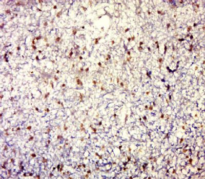
 Paraformaldehyde-fixed, paraffin embedded (Human brain glioma); Antigen retrieval by boiling in sodium citrate buffer (pH6.0) for 15min; Block endogenous peroxidase by 3% hydrogen peroxide for 20 minutes; Blocking buffer (normal goat serum) at 37°C for 30min; Antibody incubation with (MUC1) Polyclonal Antibody, Unconjugated (bs-1018R) at 1:500 overnight at 4°C, followed by a conjugated secondary (sp-0023) for 20 minutes and DAB staining.
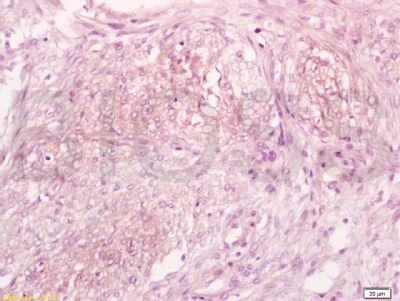
 Tissue/cell: human breast tissue; 4% Paraformaldehyde-fixed and paraffin-embedded;
Antigen retrieval: citrate buffer ( 0.01M, pH 6.0 ), Boiling bathing for 15min; Block endogenous peroxidase by 3% Hydrogen peroxide for 30min; Blocking buffer (normal goat serum,C-0005) at 37℃ for 20 min;
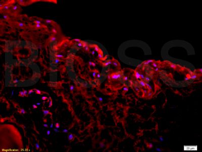
Incubation: Anti-CA15-3/Muc1 Polyclonal Antibody, Unconjugated(bs-1018R) 1:200, overnight at 4°C, followed by conjugation to the secondary antibody(SP-0023) and DAB(C-0010) staining Tissue/cell: rabbit bladder;4% Paraformaldehyde-fixed and paraffin-embedded
Antigen retrieval: citrate buffer ( 0.01M, pH 6.0 ), Boiling bathing for 15min; Blocking buffer (normal goat serum) at 37℃ for 20 min
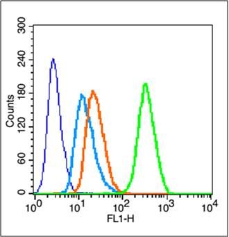
Incubation: Anti-CA15-3/Muc1 Polyclonal Antibody, Unconjugated(bs-1018R) 1:200, overnight at 4°C, The secondary antibody was Goat Anti-Rabbit IgG, PE conjugated(bs-0295G-PE)used at 1:200 dilution for 40 minutes at 37°C. DAPI(5ug/ml,blue) was used to stain the cell nuclei Blank control (blue line): MCF7
Primary Antibody (green line): Rabbit Anti-UC1 antibody (bs-1018R)
Dilution: 1μg /10^6 cells;
Isotype Control Antibody (orange line): Rabbit IgG .
Secondary Antibody (white blue line): Goat anti-rabbit IgG-FITC
Dilution: 1μg /test.
Protocol
The cells were fixed with 70% ethanol (Overnight at 4℃) and then permeabilized with 90% ice-cold methanol for 30 min on ice. Cells stained with Primary Antibody for 30 min at room temperature. The cells were then incubated in 1 X PBS/2%BSA/10% goat serum to block non-specific protein-protein interactions followed by the antibody for 15 min at room temperature. The secondary antibody used for 40 min at room temperature. Acquisition of 20,000 events was performed.

