产品中心
当前位置:首页>产品中心Anti-protein C
货号: bs-0040R 基本售价: 1380.0 元 规格: 100ul
- 规格:100ul
- 价格:1380.00元
- 规格:200ul
- 价格:2200.00元
产品信息
- 产品编号
- bs-0040R
- 英文名称
- protein C
- 中文名称
- 维生素K依赖的蛋白C重链抗体
- 别 名
- Anticoagulant protein C; Autoprothrombin IIA; Blood coagulation factor XIV; EC 3.4.21.69; PC; PROC; PROC1; Vitamin K dependent protein C precursor; APC; EC 3.4.21.69; PC; proC; PROC_HUMAN; Protein C (inactivator of coagulation factors Va and VIIIa); Vitamin K dependent protein C; Vitamin K-dependent protein C; Anticoagulant protein C; Vitamin K-dependent protein C heavy chain.
- 规格价格
- 100ul/1380元购买 200ul/2200元购买 大包装/询价
- 说 明 书
- 100ul 200ul
- 研究领域
- 心血管 细胞生物 免疫学 信号转导
- 抗体来源
- Rabbit
- 克隆类型
- Polyclonal
- 交叉反应
- Human, Mouse, Rat, Chicken, Dog, Pig, Cow,
- 产品应用
- WB=1:500-2000 ELISA=1:500-1000 IHC-P=1:400-800 IHC-F=1:400-800 IF=1:100-500 (石蜡切片需做抗原修复)
not yet tested in other applications.
optimal dilutions/concentrations should be determined by the end user.
- 分 子 量
- 29/46kDa
- 细胞定位
- 分泌型蛋白
- 性 状
- Lyophilized or Liquid
- 浓 度
- 1mg/ml
- 免 疫 原
- KLH conjugated synthetic peptide derived from human Vitamin K-dependent protein C heavy chain:371-460/461
- 亚 型
- IgG
- 纯化方法
- affinity purified by Protein A
- 储 存 液
- 0.01M TBS(pH7.4) with 1% BSA, 0.03% Proclin300 and 50% Glycerol.
- 保存条件
- Store at -20 °C for one year. Avoid repeated freeze/thaw cycles. The lyophilized antibody is stable at room temperature for at least one month and for greater than a year when kept at -20°C. When reconstituted in sterile pH 7.4 0.01M PBS or diluent of antibody the antibody is stable for at least two weeks at 2-4 °C.
- PubMed
- PubMed
- 产品介绍
- background:
This gene encodes a vitamin K-dependent plasma glycoprotein. The encoded protein is cleaved to its activated form by the thrombin-thrombomodulin complex. This activated form contains a serine protease domain and functions in degradation of the activated forms of coagulation factors V and VIII. Mutations in this gene have been associated with thrombophilia due to protein C deficiency, neonatal purpura fulminans, and recurrent venous thrombosis.[provided by RefSeq, Dec 2009].
Function:
Protein C is a vitamin K-dependent serine protease that regulates blood coagulation by inactivating factors Va and VIIIa in the presence of calcium ions and phospholipids.
Subunit:
Synthesized as a single chain precursor, which is cleaved into a light chain and a heavy chain held together by a disulfide bond. The enzyme is then activated by thrombin, which cleaves a tetradecapeptide from the amino end of the heavy chain; this reaction, which occurs at the surface of endothelial cells, is strongly promoted by thrombomodulin.
Tissue Specificity:
Plasma; synthesized in the liver.
Post-translational modifications:
The vitamin K-dependent, enzymatic carboxylation of some Glu residues allows the modified protein to bind calcium.
N- and O-glycosylated. Partial (70%) N-glycosylation of Asn-371 with an atypical N-X-C site produces a higher molecular weight form referred to as alpha. The lower molecular weight form, not N-glycosylated at Asn-371, is beta. O-glycosylated with core 1 or possibly core 8 glycans.
The iron and 2-oxoglutarate dependent 3-hydroxylation of aspartate and asparagine is (R) stereospecific within EGF domains.
May be phosphorylated on a Ser or Thr in a region (AA 25-30) of the propeptide.
DISEASE:
Defects in PROC are the cause of thrombophilia due to protein C deficiency, autosomal dominant (THPH3) [MIM:176860]. A hemostatic disorder characterized by impaired regulation of blood coagulation and a tendency to recurrent venous thrombosis. However, many adults with heterozygous disease may be asymptomatic. Individuals with decreased amounts of protein C are classically referred to as having type I protein C deficiency and those with normal amounts of a functionally defective protein as having type II deficiency.
Defects in PROC are the cause of thrombophilia due to protein C deficiency, autosomal recessive (THPH4) [MIM:612304]. A hemostatic disorder characterized by impaired regulation of blood coagulation and a tendency to recurrent venous thrombosis. It results in a thrombotic condition that can manifest as a severe neonatal disorder or as a milder disorder with late-onset thrombophilia. The severe form leads to neonatal death through massive neonatal venous thrombosis. Often associated with ecchymotic skin lesions which can turn necrotic called purpura fulminans, this disorder is very rare.
Similarity:
Belongs to the peptidase S1 family.
Contains 2 EGF-like domains.
Contains 1 Gla (gamma-carboxy-glutamate) domain.
Contains 1 peptidase S1 domain.
SWISS:
P04070
Gene ID:
5624
Database links:Entrez Gene: 5624 Human
Omim: 612283 Human
SwissProt: P04070 Human
Unigene: 224698 Human
Important Note:
This product as supplied is intended for research use only, not for use in human, therapeutic or diagnostic applications.
活化蛋白C是一种丝氨酸蛋白酶,也是一种抑癌基因,参与细胞信号的传导,在细胞分裂、细胞黏附中有重要的作用
有人用于抑制凝血 (抗凝作用)促进纤维蛋白溶解及抗炎作用的研究, 近年来有学者认为APC还可以抑制血管内皮细胞凋亡, 有抑制肿瘤坏死因子产生、限制凝血酶诱导炎症反应与微血管内皮细胞的一些作用。
- 产品图片
 Sample:
Hcclm3(Human) Cell Lysate at 30 ug
Primary: Anti-protein C (bs-0040R) at 1/1000 dilution
Secondary: IRDye800CW Goat Anti-Rabbit IgG at 1/20000 dilution
Predicted band size: 29/46 kD
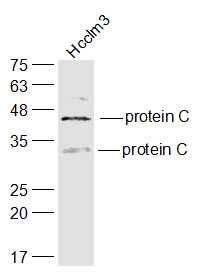
Observed band size: 29/46 kD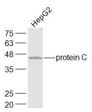 Sample:
HepG2(Human) Cell Lysate at 30 ug
Primary: Anti-protein C (bs-0040R) at 1/1000 dilution
Secondary: IRDye800CW Goat Anti-Rabbit IgG at 1/20000 dilution
Predicted band size: 29/46 kD
Observed band size: 46 kD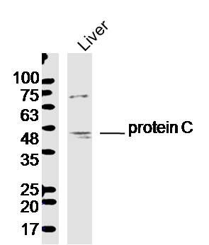 Sample: Liver (Mouse) Lysate at 40 ug
Primary: Anti-protein C (bs-0040R) at 1/300 dilution
Secondary: IRDye800CW Goat Anti-Rabbit IgG at 1/20000 dilution
Predicted band size: 29/46 kD
Observed band size: 46 kD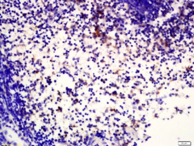 Tissue/cell: Mouse spleen tissue; 4% Paraformaldehyde-fixed and paraffin-embedded;
Antigen retrieval: citrate buffer ( 0.01M, pH 6.0 ), Boiling bathing for 15min; Block endogenous peroxidase by 3% Hydrogen peroxide for 30min; Blocking buffer (normal goat serum,C-0005) at 37℃ for 20 min;
Incubation: Anti-protein C Polyclonal Antibody, Unconjugated(bs-0040R) 1:200, overnight at 4°C, followed by conjugation to the secondary antibody(SP-0023) and DAB(C-0010) staining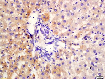 Tissue/cell: Rat liver tissue; 4% Paraformaldehyde-fixed and paraffin-embedded;
Antigen retrieval: citrate buffer ( 0.01M, pH 6.0 ), Boiling bathing for 15min; Block endogenous peroxidase by 3% Hydrogen peroxide for 30min; Blocking buffer (normal goat serum,C-0005) at 37℃ for 20 min;
Incubation: Anti-protein C Polyclonal Antibody, Unconjugated(bs-0040R) 1:200, overnight at 4°C, followed by conjugation to the secondary antibody(SP-0023) and DAB(C-0010) staining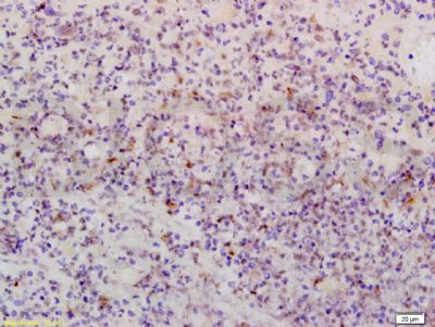 Tissue/cell: Human brain glioma tissue; 4% Paraformaldehyde-fixed and paraffin-embedded;
Antigen retrieval: citrate buffer ( 0.01M, pH 6.0 ), Boiling bathing for 15min; Block endogenous peroxidase by 3% Hydrogen peroxide for 30min; Blocking buffer (normal goat serum,C-0005) at 37℃ for 20 min;
Incubation: Anti-Activated protein C/PROC1 Polyclonal Antibody, Unconjugated(bs-0040R) 1:200, overnight at 4°C, followed by conjugation to the secondary antibody(SP-0023) and DAB(C-0010) staining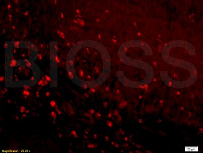 Tissue/cell: rat colitis tissue;4% Paraformaldehyde-fixed and paraffin-embedded;
Antigen retrieval: citrate buffer ( 0.01M, pH 6.0 ), Boiling bathing for 15min; Blocking buffer (normal goat serum,C-0005) at 37℃ for 20 min;
Incubation: Anti-Activated protein C/PROC1 Polyclonal Antibody, Unconjugated(bs-0040R) 1:200, overnight at 4°C; The secondary antibody was Goat Anti-Rabbit IgG, PE conjugated (bs-0295G-PE)used at 1:200 dilution for 40 minutes at 37°C.

