产品中心
当前位置:首页>产品中心Anti-PIWIL1
货号: bs-20224R 基本售价: 1380.0 元 规格: 100ul
- 规格:100ul
- 价格:1380.00元
- 规格:200ul
- 价格:2200.00元
产品信息
- 产品编号
- bs-20224R
- 英文名称
- PIWIL1
- 中文名称
- piwi样1蛋白抗体
- 别 名
- PIWL1_HUMAN; Piwi-like protein 1.
- 规格价格
- 100ul/1380元购买 200ul/2200元购买 大包装/询价
- 说 明 书
- 100ul 200ul
- 研究领域
- 肿瘤 细胞生物 免疫学 信号转导 转录调节因子
- 抗体来源
- Rabbit
- 克隆类型
- Polyclonal
- 交叉反应
- Human, Mouse, Chicken, Dog, Pig, Cow, Horse, Rabbit, Sheep,
- 产品应用
- WB=1:500-2000 ELISA=1:500-1000 IHC-P=1:400-800 IHC-F=1:400-800 Flow-Cyt=1μg/Test ICC=1:100-500 IF=1:100-500 (石蜡切片需做抗原修复)
not yet tested in other applications.
optimal dilutions/concentrations should be determined by the end user.
- 分 子 量
- 99kDa
- 细胞定位
- 细胞浆
- 性 状
- Lyophilized or Liquid
- 浓 度
- 1mg/ml
- 免 疫 原
- KLH conjugated synthetic peptide derived from human PIWIL1:421-520/861
- 亚 型
- IgG
- 纯化方法
- affinity purified by Protein A
- 储 存 液
- 0.01M TBS(pH7.4) with 1% BSA, 0.03% Proclin300 and 50% Glycerol.
- 保存条件
- Store at -20 °C for one year. Avoid repeated freeze/thaw cycles. The lyophilized antibody is stable at room temperature for at least one month and for greater than a year when kept at -20°C. When reconstituted in sterile pH 7.4 0.01M PBS or diluent of antibody the antibody is stable for at least two weeks at 2-4 °C.
- PubMed
- PubMed
- 产品介绍
- Function:
1 attaches the virion to the cell membrane by interacting with human ACE2 and CLEC4M/DC-SIGNR, initiating the infection. Binding to the receptor and internalization of the virus into the endosomes of the host cell probably induces conformational changes in the S glycoprotein. Proteolysis by cathepsin CTSL may unmask the fusion peptide of S2 and activate membranes fusion within endosomes.
S2 is a class I viral fusion protein. Under the current model, the protein has at least three conformational states: pre-fusion native state, pre-hairpin intermediate state, and post-fusion hairpin state. During viral and target cell membrane fusion, the coiled coil regions (heptad repeats) assume a trimer-of-hairpins structure, positioning the fusion peptide in close proximity to the C-terminal region of the ectodomain. The formation of this structure appears to drive apposition and subsequent fusion of viral and target cell membranes.
Subunit:
Homotrimer. Binds to human and palm civet ACE2 and human CLEC4M/DC-SIGNR. Interacts with the accessory proteins 3a and 7a. {ECO:0000269|PubMed:14647384, ECO:0000269|PubMed:14670965, ECO:0000269|PubMed:15194747, ECO:0000269|PubMed:15496474, ECO:0000269|PubMed:16166518, ECO:0000269|PubMed:16840309}.
Subcellular Location:
Virion membrane {ECO:0000269|PubMed:15831954}; Single-pass type I membrane protein {ECO:0000269|PubMed:15831954}. Host endoplasmic reticulum-Golgi intermediate compartment membrane {ECO:0000250}; Single-pass type I membrane protein {ECO:0000250}. Host cell membrane {ECO:0000269|PubMed:15831954}; Single-pass type I membrane protein {ECO:0000269|PubMed:15831954}. Note=Accumulates in the endoplasmic reticulum-Golgi intermediate compartment, where it participates in virus particle assembly (By similarity). Some S oligomers are transported to the plasma membrane, where they may mediate cell-cell fusion. {ECO:0000250}.
Post-translational modifications:
he cytoplasmic Cys-rich domain is palmitoylated. Spike glycoprotein is digested by cathepsin CTSL within endosomes. {ECO:0000269|PubMed:17134730}.
Similarity:
Belongs to the coronaviruses spike protein family. {ECO:0000305}.
SWISS:
Q96J94
Gene ID:
9271
Database links:Entrez Gene: 9271 Human
Entrez Gene: 57749 Mouse
Entrez Gene: 363912 Rat
Omim: 605571 Human
SwissProt: Q96J94 Human
SwissProt: Q9JMB7 Mouse
Unigene: 405659 Human
Unigene: 272720 Mouse
Unigene: 131387 Rat
Important Note:
This product as supplied is intended for research use only, not for use in human, therapeutic or diagnostic applications.
- 产品图片
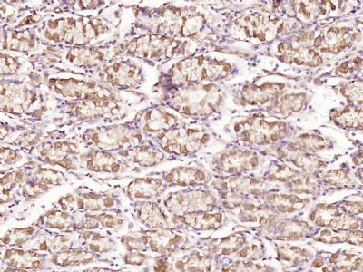
 Paraformaldehyde-fixed, paraffin embedded (Human stomach carcinoma); Antigen retrieval by boiling in sodium citrate buffer (pH6.0) for 15min; Block endogenous peroxidase by 3% hydrogen peroxide for 20 minutes; Blocking buffer (normal goat serum) at 37°C for 30min; Antibody incubation with (PIWL1) Polyclonal Antibody, Unconjugated (bs-20224R) at 1:500 overnight at 4°C, followed by a conjugated secondary (sp-0023) for 20 minutes and DAB staining.
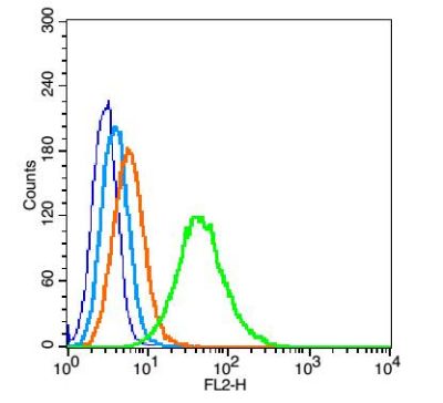
 Blank control(blue): TM4 cells(fixed with 2% paraformaldehyde (10 min) , then permeabilized with 90% ice-cold methanol for 30 min on ice).
Primary Antibody:Rabbit Anti-PIWIL1 antibody(bs-20224R), Dilution: 5μg in 100 μL 1X PBS containing 0.5% BSA;
Isotype Control Antibody: Rabbit IgG(orange) ,used under the same conditions );
Secondary Antibody: Goat anti-rabbit IgG-PE(white blue), Dilution: 1:200 in 1 X PBS containing 0.5% BSA.

