产品中心
当前位置:首页>产品中心Anti-MUL1
货号: bs-9291R 基本售价: 1380.0 元 规格: 100ul
- 规格:100ul
- 价格:1380.00元
- 规格:200ul
- 价格:2200.00元
产品信息
- 产品编号
- bs-9291R
- 英文名称
- MUL1
- 中文名称
- E3泛素连接酶MUL1抗体
- 别 名
- E3 ubiquitin-protein ligase MUL1; C1orf166; E3 ubiquitin ligase; E3 ubiquitin protein ligase MUL1; GIDE; Growth inhibition and death E3 ligase; MAPL; Mitochondrial anchored protein ligase; Mitochondrial ubiquitin ligase activator of NFKB 1; MUL1; MULAN; Putative NF kappa B activating protein 266; RING finger protein 218; RNF218; RP23-25C1.10-002; MUL1_HUMAN.
- 规格价格
- 100ul/1380元购买 200ul/2200元购买 大包装/询价
- 说 明 书
- 100ul 200ul
- 研究领域
- 细胞生物 免疫学
- 抗体来源
- Rabbit
- 克隆类型
- Polyclonal
- 交叉反应
- Human, Mouse, Rat, Dog, Pig, Cow, Rabbit,
- 产品应用
- WB=1:500-2000 ELISA=1:500-1000 IHC-P=1:400-800 IHC-F=1:400-800 Flow-Cyt=1ug/test IF=1:50-200 (石蜡切片需做抗原修复)
not yet tested in other applications.
optimal dilutions/concentrations should be determined by the end user.
- 分 子 量
- 40kDa
- 细胞定位
- 细胞浆 细胞膜
- 性 状
- Lyophilized or Liquid
- 浓 度
- 1mg/ml
- 免 疫 原
- KLH conjugated synthetic peptide derived from human MUL1/RNF218:1-100/352
- 亚 型
- IgG
- 纯化方法
- affinity purified by Protein A
- 储 存 液
- 0.01M TBS(pH7.4) with 1% BSA, 0.03% Proclin300 and 50% Glycerol.
- 保存条件
- Store at -20 °C for one year. Avoid repeated freeze/thaw cycles. The lyophilized antibody is stable at room temperature for at least one month and for greater than a year when kept at -20°C. When reconstituted in sterile pH 7.4 0.01M PBS or diluent of antibody the antibody is stable for at least two weeks at 2-4 °C.
- PubMed
- PubMed
- 产品介绍
- background:
E3 ubiquitin-protein ligase that plays a role in the control of mitochondrial morphology. Promotes mitochondrial fragmentation and influences mitochondrial localization. Inhibits cell growth. When overexpressed, activates JNK through MAP3K7/TAK1 and induces caspase-dependent apoptosis. E3 ubiquitin ligases accept ubiquitin from an E2 ubiquitin-conjugatin.
Function:
Exhibits weak E3 ubiquitin-protein ligase activity, but preferentially acts as a SUMO E3 ligase at physiological concentrations. Plays a role in the control of mitochondrial morphology. Promotes mitochondrial fragmentation and influences mitochondrial localization. Inhibits cell growth. When overexpressed, activates JNK through MAP3K7/TAK1 and induces caspase-dependent apoptosis. E3 ubiquitin ligases accept ubiquitin from an E2 ubiquitin-conjugating enzyme in the form of a thioester and then directly transfer the ubiquitin to targeted substrates.
Subunit:
Homooligomer. Interacts with MAP3K7/TAK1. Interacts with UBC9. Interacts with and sumoylates DNM1L.
Subcellular Location:
Mitochondrion outer membrane; Multi-pass membrane protein. Peroxisome. Note: Transported in mitochondrion-derived vesicles from the mitochondrion to the peroxisome.
Tissue Specificity:
Widely expressed with highest levels in the heart, skeletal muscle, placenta, kidney and liver. Barely detectable in colon and thymus.
Similarity:
Contains 1 RING-type zinc finger.
SWISS:
Q969V5
Gene ID:
79594
Database links:Entrez Gene: 79594 Human
Entrez Gene: 68350 Mouse
Entrez Gene: 298576 Rat
Omim: 612037 Human
SwissProt: Q969V5 Human
SwissProt: Q8VCM5 Mouse
Unigene: 10101 Human
Unigene: 103413 Mouse
Important Note:
This product as supplied is intended for research use only, not for use in human, therapeutic or diagnostic applications.
- 产品图片
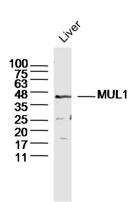 Sample: Liver (Mouse) Lysate at 40 ug
Primary: Anti-MUL1 (bs-9291R)at 1/300 dilution
Secondary: IRDye800CW Goat Anti-Rabbit IgG at 1/20000 dilution
Predicted band size: 40kD
Observed band size: 40kD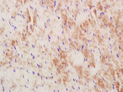 Paraformaldehyde-fixed, paraffin embedded (Rat skeletal muscle); Antigen retrieval by boiling in sodium citrate buffer (pH6.0) for 15min; Block endogenous peroxidase by 3% hydrogen peroxide for 20 minutes; Blocking buffer (normal goat serum) at 37°C for 30min; Antibody incubation with (MUL1) Polyclonal Antibody, Unconjugated (bs-9291R) at 1:400 overnight at 4°C, followed by a conjugated secondary antibody (sp-0023) for 20 minutes and DAB staining.
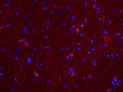 Paraformaldehyde-fixed, paraffin embedded (Mouse brain); Antigen retrieval by boiling in sodium citrate buffer (pH6.0) for 15min; Block endogenous peroxidase by 3% hydrogen peroxide for 20 minutes; Blocking buffer (normal goat serum) at 37°C for 30min; Antibody incubation with (MUL1) Polyclonal Antibody, Unconjugated (bs-9291R) at 1:400 overnight at 4°C, followed by a conjugated secondary antibody (bs-0295g-cy3) for 90 minutes, and DAPI for nuclei staining.
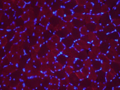 Paraformaldehyde-fixed, paraffin embedded (Rat skeletal muscle); Antigen retrieval by boiling in sodium citrate buffer (pH6.0) for 15min; Block endogenous peroxidase by 3% hydrogen peroxide for 20 minutes; Blocking buffer (normal goat serum) at 37°C for 30min; Antibody incubation with (MUL1) Polyclonal Antibody, Unconjugated (bs-9291R) at 1:400 overnight at 4°C, followed by a conjugated secondary antibody (bs-0295g-cy3) for 90 minutes, and DAPI for nuclei staining.
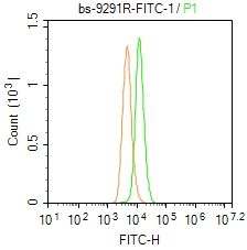 Blank control: A549.
Primary Antibody (green line): Rabbit Anti-MUL1/FITC Conjugated antibody (bs-9291R-FITC)
Dilution: 1μg /10^6 cells;
Isotype Control Antibody (orange line): Rabbit IgG-FITC .
Protocol
The cells were fixed with 4% PFA (10min at room temperature)and then permeabilized with 0.1% PBST for 20 min at-20℃. The cells were then incubated in 5% BSA to block non-specific protein-protein interactions for 30 min at room temperature. The cells were stained with Primary Antibody for 30 min at room temperature. Acquisition of 20,000 events was performed.

