产品中心
当前位置:首页>产品中心Anti-P4HB
货号: bs-1476R 基本售价: 780.0 元 规格: 50ul
- 规格:50ul
- 价格:780.00元
- 规格:100ul
- 价格:1380.00元
- 规格:200ul
- 价格:2200.00元
产品信息
- 产品编号
- bs-1476R
- 英文名称
- P4HB
- 中文名称
- 蛋白质二硫键异构酶抗体
- 别 名
- Cellular thyroid hormone binding protein; Cellular thyroid hormone-binding protein; Collagen prolyl 4 hydroxylase beta; Disulphide Isomerase; DSI; EC 5.3.4.1; Endoplasmic reticulum resident protein 59; ER protein 59; ERBA2L; ERp59; GIT; Gltathione insulin transhydrogenase; Glutathione insulin transhydrogenase; P4Hbeta; p55; PDI; PDIA1; PDIA1_HUMAN; PDIR; PHDB; PO4DB; PO4HB; Procollagen proline 2 oxoglutarate 4 dioxygenase (proline 4 hydroxylase) beta polypeptide (protein disulfide isomerase associated 1); Procollagen proline 2 oxoglutarate 4 dioxygenase beta subunit; PROHB; Prolyl 4 hydroxylase beta polypeptide; Prolyl 4 hydroxylase beta subunit; Prolyl 4 hydroxylase subunit beta; Prolyl 4-hydroxylase subunit beta; Protein disulfide isomerase associated 1; Protein disulfide isomerase, family A, member 1; Protein disulfide isomerase/oxidoreductase; Protein disulfide-isomerase; Protocollagen hydroxylase; Thbp; Thyroid hormone binding protein p55; Thyroid hormone binding protein p55 cellular; V erb a avian erythroblastic leukemia viral oncogene homolog 2 like.
- 规格价格
- 50ul/780元购买 100ul/1380元购买 200ul/2200元购买 大包装/询价
- 说 明 书
- 50ul 100ul 200ul
- 研究领域
- 细胞生物 信号转导 激酶和磷酸酶
- 抗体来源
- Rabbit
- 克隆类型
- Polyclonal
- 交叉反应
- Human, Mouse, Rat, Chicken, Cow, Horse, Rabbit,
- 产品应用
- WB=1:500-2000 ELISA=1:500-1000 IHC-P=1:400-800 IHC-F=1:400-800 IF=1:100-500 (石蜡切片需做抗原修复)
not yet tested in other applications.
optimal dilutions/concentrations should be determined by the end user.
- 分 子 量
- 55kDa
- 细胞定位
- 细胞浆
- 性 状
- Lyophilized or Liquid
- 浓 度
- 1mg/ml
- 免 疫 原
- KLH conjugated synthetic peptide derived from human PDI:21-150/508
- 亚 型
- IgG
- 纯化方法
- affinity purified by Protein A
- 储 存 液
- 0.01M TBS(pH7.4) with 1% BSA, 0.03% Proclin300 and 50% Glycerol.
- 保存条件
- Store at -20 °C for one year. Avoid repeated freeze/thaw cycles. The lyophilized antibody is stable at room temperature for at least one month and for greater than a year when kept at -20°C. When reconstituted in sterile pH 7.4 0.01M PBS or diluent of antibody the antibody is stable for at least two weeks at 2-4 °C.
- PubMed
- PubMed
- 产品介绍
- background:
The three dimensional structure of many extracellular proteins is stabilized by the formation of disulphide bonds. Studies suggest that a microsomal enzyme known as Protein Disulphide Isomerase (PDI) is involved in disulphide-bond formation and isomerization, as well as the reduction of disulphide bonds in proteins. PDI, which catalyses disulphide interchange between thiols and protein dilsulphides, has also been referred to as thiol:protein-disulphide oxidoreductase and as glutathione:insulin transhydrogenase because of its role in reduction of disulphide bonds. The highly conserved sequence Lys-Asp-Glu-Leu (KDEL) is present at the carboxy-terminus of PDI and other soluble endoplasmic reticulum (ER) resident proteins including the 78 and 94 kDa glucose regulated proteins (GRP78 and GRP94 respectively). The presence of carboxy-terminal KDEL appears to be necessary for ER retention and appears to be sufficient to reduce the secretion of proteins from the ER. This retention is reported to be mediated by a KDEL receptor.
Function:
This multifunctional protein catalyzes the formation, breakage and rearrangement of disulfide bonds. At the cell surface, seems to act as a reductase that cleaves disulfide bonds of proteins attached to the cell. May therefore cause structural modifications of exofacial proteins. Inside the cell, seems to form/rearrange disulfide bonds of nascent proteins. At high concentrations, functions as a chaperone that inhibits aggregation of misfolded proteins. At low concentrations, facilitates aggregation (anti-chaperone activity). May be involved with other chaperones in the structural modification of the TG precursor in hormone biogenesis. Also acts a structural subunit of various enzymes such as prolyl 4-hydroxylase and microsomal triacylglycerol transfer protein MTTP.
Subunit:
Homodimer. Monomers and homotetramers may also occur. Also constitutes the structural subunit of prolyl 4-hydroxylase and of the microsomal triacylglycerol transfer protein MTTP in mammalian cells. Stabilizes both enzymes and retain them in the ER without contributing to the catalytic activity (By similarity). Binds UBQLN1. Binds to CD4, and upon HIV-1 binding to the cell membrane, is part of a P4HB/PDI-CD4-CXCR4-gp120 complex.
Subcellular Location:
Endoplasmic reticulum lumen. Melanosome. Cell membrane; Peripheral membrane protein (Potential). Note=Highly abundant. In some cell types, seems to be also secreted or associated with the plasma membrane, where it undergoes constant shedding and replacement from intracellular sources (Probable). Localizes near CD4-enriched regions on lymphoid cell surfaces. Identified by mass spectrometry in melanosome fractions from stage I to stage IV.
Tissue Specificity:
Detected in the flagellum and head region of spermatozoa (at protein level).
Similarity:
Belongs to the protein disulfide isomerase family.
Contains 2 thioredoxin domains.
SWISS:
P07237
Gene ID:
5034
Database links:Entrez Gene: 5034 Human
Entrez Gene: 18453 Mouse
Entrez Gene: 25506 Rat
Omim: 176790 Human
SwissProt: P07237 Human
SwissProt: P09103 Mouse
SwissProt: P04785 Rat
Unigene: 464336 Human
Unigene: 708779 Human
Unigene: 16660 Mouse
Unigene: 392196 Mouse
Unigene: 4234 Rat
Important Note:
This product as supplied is intended for research use only, not for use in human, therapeutic or diagnostic applications.
蛋白质二硫键异构酶(PDI,Protein Disulfide lsomerase)是一种亚基分子量为57kD的二聚体蛋白,一般认为它参与细胞内蛋白质天然二硫键的形成,它催化蛋白质巯基和二硫键的交换反应.
- 产品图片
 Sample:
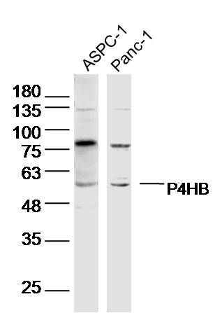
ASPC-1 Cell (Human) Lysate at 40 ug
PANC-1 Cell (Human) Lysate at 40 ug
Primary: Anti- P4HB (bs-1476R) at 1/300 dilution
Secondary: IRDye800CW Goat Anti-Rabbit IgG at 1/20000 dilution
Predicted band size: 55 kD
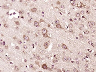
Observed band size: 55 kD Paraformaldehyde-fixed, paraffin embedded (Mouse brain); Antigen retrieval by boiling in sodium citrate buffer (pH6.0) for 15min; Block endogenous peroxidase by 3% hydrogen peroxide for 20 minutes; Blocking buffer (normal goat serum) at 37°C for 30min; Antibody incubation with (P4HB) Polyclonal Antibody, Unconjugated (bs-1476R) at 1:400 overnight at 4°C, followed by a conjugated secondary antibody (sp-0023) for 20 minutes and DAB staining.

