产品中心
当前位置:首页>产品中心Anti-RIPK-1
货号: bs-5805R 基本售价: 1380.0 元 规格: 100ul
- 规格:100ul
- 价格:1380.00元
- 规格:200ul
- 价格:2200.00元
产品信息
- 产品编号
- bs-5805R
- 英文名称
- RIPK-1
- 中文名称
- 丝氨酸苏氨酸激酶1受体相互作用蛋白抗体
- 别 名
- Cell death protein RIP; Receptor (TNFRSF) interacting serine threonine kinase 1; Receptor interacting protein; Receptor interacting serine threonine protein kinase 1; Receptor TNFRSF interacting serine threonine kinase 1; Receptor-interacting protein 1; Receptor-interacting serine/threonine-protein kinase 1; Rinp; RIP; Rip-1; RIPK 1; Ripk1; RIPK1_HUMAN; Serine threonine protein kinase RIP; Serine/threonine-protein kinase RIP.

- Specific References (1) | bs-5805R has been referenced in 1 publications.[IF=2.84] Luo, Yuhao, et al. "Lycorine induces programmed necrosis in the multiple myeloma cell line ARH-77." Tumor Biology (2014): 1-9. WB ; Human.PubMed:25487618
- 规格价格
- 100ul/1380元购买 200ul/2200元购买 大包装/询价
- 说 明 书
- 100ul 200ul
- 研究领域
- 肿瘤 细胞生物 免疫学 细胞凋亡 激酶和磷酸酶
- 抗体来源
- Rabbit
- 克隆类型
- Polyclonal
- 交叉反应
- Human, Mouse, Rat, Pig, Cow, Horse, Rabbit,
- 产品应用
- WB=1:500-2000 ELISA=1:500-1000 IHC-P=1:400-800 IHC-F=1:400-800 Flow-Cyt=2ug/Test IF=1:100-500 (石蜡切片需做抗原修复)
not yet tested in other applications.
optimal dilutions/concentrations should be determined by the end user.
- 分 子 量
- 74kDa
- 细胞定位
- 细胞浆 细胞膜
- 性 状
- Lyophilized or Liquid
- 浓 度
- 1mg/ml
- 免 疫 原
- KLH conjugated synthetic peptide derived from human RIPK-1/RIP:581-671/671
- 亚 型
- IgG
- 纯化方法
- affinity purified by Protein A
- 储 存 液
- 0.01M TBS(pH7.4) with 1% BSA, 0.03% Proclin300 and 50% Glycerol.
- 保存条件
- Store at -20 °C for one year. Avoid repeated freeze/thaw cycles. The lyophilized antibody is stable at room temperature for at least one month and for greater than a year when kept at -20°C. When reconstituted in sterile pH 7.4 0.01M PBS or diluent of antibody the antibody is stable for at least two weeks at 2-4 °C.
- PubMed
- PubMed
- 产品介绍
- background:
Essential adapter molecule for the activation of NF-kappa-B. Following different upstream signals (binding of inflammatory cytokines, stimulation of pathogen recognition receptors, or DNA damage), particular RIPK1-containing complexes are formed, initiating a limited number of cellular responses. Upon TNFA stimulation RIPK1 is recruited to a TRADD-TRAF complex initiated by TNFR1 trimerization. There, it is ubiquitinated via Lys-63-link chains, inducing its association with the IKK complex, and its activation through NEMO binding of polyubiquitin chains.
Function:
Serine-threonine kinase which transduces inflammatory and cell-death signals (necroptosis) following death receptors ligation, activation of pathogen recognition receptors (PRRs), and DNA damage. Upon activation of TNFR1 by the TNF-alpha family cytokines, TRADD and TRAF2 are recruited to the receptor. Ubiquitination by TRAF2 via Lys-63-link chains acts as a critical enhancer of communication with downstream signal transducers in the mitogen-activated protein kinase pathway and the NF-kappa-B pathway, which in turn mediate downstream events including the activation of genes encoding inflammatory molecules. Polyubiquitinated protein binds to IKBKG/NEMO, the regulatory subunit of the IKK complex, a critical event for NF-kappa-B activation. Interaction with other cellular RHIM-containing adapters initiates gene activation and cell death. RIPK1 and RIPK3 association, in particular, forms a necroptosis-inducing complex.
Subunit:
Interacts (via RIP homotypic interaction motif) with RIPK3 (via RIP homotypic interaction motif); this interaction induces RIPK1 necroptosis-specific phosphorylation, formation of the necroptosis-inducing complex. Interacts (via the death domain) with TNFRSF6 (via the death domain) and TRADD (via the death domain). Is recruited by TRADD to TNFRSF1A in a TNF-dependent process. Binds RNF216, EGFR, IKBKG, TRAF1, TRAF2 and TRAF3. Interacts with BNLF1. Interacts with SQSTM1 upon TNF-alpha stimulation. May interact with MAVS/IPS1. Interacts with ZFAND5. Interacts with RBCK1 (By similarity).
Subcellular Location:
Cytoplasm. Cell membrane (By similarity).
Post-translational modifications:
Proteolytically cleaved by caspase-8 during TNF-induced apoptosis. Cleavage abolishes NF-kappa-B activation and enhances pro-apototic signaling through the TRADD-FADD interaction.
RIPK1 and RIPK3 undergo reciprocal auto- and trans-phosphorylation. Phosphorylation of Ser-161 by RIPK3 is necessary for the formation of the necroptosis-inducing complex.
Ubiquitinated by Lys-11-, Lys-48-, Lys-63- and linear-linked type ubiquitin. Polyubiquitination with Lys-63-linked chains by TRAF2 induces association with the IKK complex. Deubiquitination of Lys-63-linked chains and polyubiquitination with Lys-48-linked chains by TNFAIP3 leads to RIPK1 proteasomal degradation and consequently downregulates TNF-alpha-induced NFkappa-B signaling. Linear polyubiquitinated; the head-to-tail polyubiquitination is mediated by the LUBAC complex. LPS-mediated activation of NF-kappa-B. Also ubiquitinated with Lys-11-linked chains.
Similarity:
Belongs to the protein kinase superfamily. TKL Ser/Thr protein kinase family. [SIMILARITY] Contains 1 death domain.
Contains 1 protein kinase domain.
SWISS:
Q13546
Gene ID:
8737
Database links:Entrez Gene: 8737 Human
Entrez Gene: 19766 Mouse
Entrez Gene: 306886 Rat
Omim: 603453 Human
SwissProt: Q13546 Human
SwissProt: Q60855 Mouse
Unigene: 519842 Human
Unigene: 374799 Mouse
Unigene: 7572 Rat
Important Note:
This product as supplied is intended for research use only, not for use in human, therapeutic or diagnostic applications.
- 产品图片
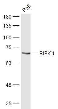 Sample:
Raji(Human) Cell Lysate at 30 ug
Primary: Anti-RIPK-1 (bs-5805R) at 1/1000 dilution
Secondary: IRDye800CW Goat Anti-Rabbit IgG at 1/20000 dilution
Predicted band size: 74 kD
Observed band size: 70 kD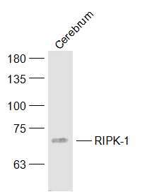 Sample:
Cerebrum (Mouse) Lysate at 40 ug
Primary: Anti-RIPK-1 (bs-5805R) at 1/1000 dilution
Secondary: IRDye800CW Goat Anti-Rabbit IgG at 1/20000 dilution
Predicted band size: 74 kD
Observed band size: 70 kD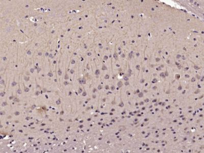 Paraformaldehyde-fixed, paraffin embedded (Mouse brain); Antigen retrieval by microwave in sodium citrate buffer (pH6.0) ; Block endogenous peroxidase by 3% hydrogen peroxide for 30 minutes; Blocking buffer (3% BSA) at RT for 30min; Antibody incubation with (RIPK-1) Polyclonal Antibody, Unconjugated (bs-5805R) at 1:400 overnight at 4℃, followed by conjugation to the secondary antibody (labeled with HRP)and DAB staining.
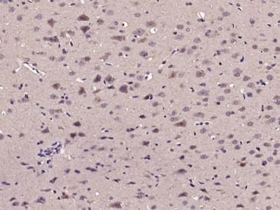 Paraformaldehyde-fixed, paraffin embedded (Rat brain); Antigen retrieval by microwave in sodium citrate buffer (pH6.0) ; Block endogenous peroxidase by 3% hydrogen peroxide for 30 minutes; Blocking buffer (3% BSA) at RT for 30min; Antibody incubation with (RIPK-1) Polyclonal Antibody, Unconjugated (bs-5805R) at 1:400 overnight at 4℃, followed by conjugation to the secondary antibody (labeled with HRP)and DAB staining.
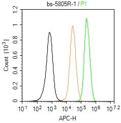 Blank control (Black line): U87MG (Black).
Primary Antibody (green line): Rabbit Anti-RIPK-1 antibody (bs-5805R)
Dilution: 1μg /10^6 cells;
Isotype Control Antibody (orange line): Rabbit IgG .
Secondary Antibody (white blue line): Goat anti-rabbit IgG-AF647
Dilution: 1μg /test.
Protocol
The cells were fixed with 4% PFA (10min at room temperature)and then permeabilized with 90% ice-cold methanol for 20 min at room temperature. The cells were then incubated in 5%BSA to block non-specific protein-protein interactions for 30 min at room temperature .Cells stained with Primary Antibody for 30 min at room temperature. The secondary antibody used for 40 min at room temperature. Acquisition of 20,000 events was performed.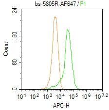 Blank control: Mouse spleen.
Primary Antibody (green line): Rabbit Anti-RIPK-1 /AF647 Conjugated antibody (bs-5805R-AF647)
Dilution: 2μg /10^6 cells;
Isotype Control Antibody (orange line): Rabbit IgG-AF647 .
Protocol
The cells were fixed with 4% PFA (10min at room temperature)and then permeabilized with 0.1% PBST for 20 min at-20℃. The cells were then incubated in 5% BSA to block non-specific protein-protein interactions for 30 min at room temperature. The cells were stained with Primary Antibody for 30 min at room temperature. Acquisition of 20,000 events was performed.

