

产品中心
当前位置:首页>产品中心Anti-CD1d
货号: bs-3690R 基本售价: 1380.0 元 规格: 100ul
- 规格:100ul
- 价格:1380.00元
- 规格:200ul
- 价格:2200.00元
产品信息
- 产品编号
- bs-3690R
- 英文名称
- CD1d
- 中文名称
- T淋巴细胞CD1D抗体
- 别 名
- Antigen-presenting glycoprotein CD1d; CD1.1; CD1d; CD1D antigen; CD1D antigen d polypeptide; CD1d molecule; CD1D_HUMAN; Cd1d1; differentiation antigen CD1 alpha 3; HMC class I antigen like glycoprotein CD1D; Ly 38; MGC34622; R3; R3G1; T cell surface glycoprotein CD1d; Thymocyte antigen CD1D.
- 规格价格
- 100ul/1380元购买 200ul/2200元购买 大包装/询价
- 说 明 书
- 100ul 200ul
- 研究领域
- 肿瘤 免疫学 信号转导 转录调节因子 细胞表面分子 t-淋巴细胞
- 抗体来源
- Rabbit
- 克隆类型
- Polyclonal
- 交叉反应
- Mouse, Rat,
- 产品应用
- WB=1:500-2000 IHC-P=1:400-800 IHC-F=1:400-800 Flow-Cyt=1μg /Test (石蜡切片需做抗原修复)
not yet tested in other applications.
optimal dilutions/concentrations should be determined by the end user.
- 分 子 量
- 36kDa
- 细胞定位
- 细胞浆 细胞膜 细胞外基质
- 性 状
- Lyophilized or Liquid
- 浓 度
- 1mg/ml
- 免 疫 原
- KLH conjugated synthetic peptide derived from mouse CD1D:51-150/336 <Extracellular>
- 亚 型
- IgG
- 纯化方法
- affinity purified by Protein A
- 储 存 液
- 0.01M TBS(pH7.4) with 1% BSA, 0.03% Proclin300 and 50% Glycerol.
- 保存条件
- Store at -20 °C for one year. Avoid repeated freeze/thaw cycles. The lyophilized antibody is stable at room temperature for at least one month and for greater than a year when kept at -20°C. When reconstituted in sterile pH 7.4 0.01M PBS or diluent of antibody the antibody is stable for at least two weeks at 2-4 °C.
- PubMed
- PubMed
- 产品介绍
- background:
This gene encodes a divergent member of the CD1 family of transmembrane glycoproteins, which are structurally related to the major histocompatibility complex (MHC) proteins and form heterodimers with beta-2-microglobulin. The CD1 proteins mediate the presentation of primarily lipid and glycolipid antigens of self or microbial origin to T cells. The human genome contains five CD1 family genes organized in a cluster on chromosome 1. The CD1 family members are thought to differ in their cellular localization and specificity for particular lipid ligands. The protein encoded by this gene localizes to late endosomes and lysosomes via a tyrosine-based motif in the cytoplasmic tail. Two transcript variants encoding different isoforms have been found for this gene. [provided by RefSeq, Jan 2016]
Function:
Antigen-presenting protein that binds self and non-self glycolipids and presents them to T-cell receptors on natural killer T-cells.
Subunit:
Heterodimer with B2M (beta-2-microglobulin). Interacts with MHC II.
Subcellular Location:
Cell membrane, Single-pass type I membrane protein. Basolateral cell membrane; Single-pass type I membrane protein. Endosome membrane; Single-pass type I membrane protein. Lysosome membrane; Single-pass type I membrane protein. Endoplasmic reticulum membrane; Single-pass type I membrane protein. Note=Subject to intracellular trafficking between the cell membrane, endosomes and lysosomes.
Tissue Specificity:
Expressed on cortical thymocytes, on certain T-cell leukemias, and in various other tissues.
Similarity:
Contains 1 Ig-like (immunoglobulin-like) domain
SWISS:
P11609
Gene ID:
12479
Database links:Entrez Gene: 12479 Mouse
Entrez Gene: 25109 Rat
SwissProt: P11609 Mouse
SwissProt: Q63493 Rat
Unigene: 1894 Mouse
Unigene: 11120 Rat
Important Note:
This product as supplied is intended for research use only, not for use in human, therapeutic or diagnostic applications.
- 产品图片
 Sample:
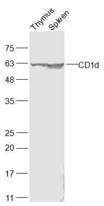
Thymus (Rat) Lysate at 40 ug
Spleen (Mouse) Lysate at 40 ug
Primary: Anti-CD1d (bs-3690R) at 1/500 dilution
Secondary: IRDye800CW Goat Anti-Rabbit IgG at 1/20000 dilution
Predicted band size: 36 kD
Observed band size: 61 kD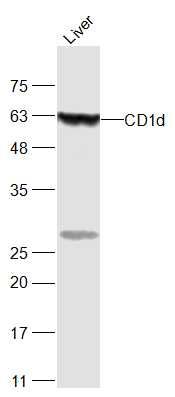 Sample:
Liver (Mouse) Lysate at 40 ug
Primary: Anti-CD1d (bs-3690R) at 1/500 dilution
Secondary: IRDye800CW Goat Anti-Rabbit IgG at 1/20000 dilution
Predicted band size: 36 kD
Observed band size: 61 kD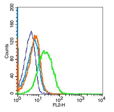 Blank control: mouse spleen cells (fixed with 2% paraformaldehyde(10 min),then permeabilized with 90% ice-cold methanol for 30 min on ice).
Primary Antibody: Rabbit Anti- CD1A antibody(bs-3690R), Dilution: 1μg in 100 μL 1X PBS containing 0.5% BSA;
Isotype Control Antibody: Rabbit IgG(orange) ,used under the same conditions );
Secondary Antibody: Goat anti-rabbit IgG-PE(white blue), Dilution: 1:200 in 1 X PBS containing 0.5% BSA.
Protocol
. Primary antibody (bs-3690R, 1μg /1x10^6 cells) were incubated for 30 min on the ice, followed by 1 X PBS containing 0.5% BSA + 1 0% goat serum (15 min) to block non-specific protein-protein interactions. Then the Goat Anti-rabbit IgG/PE antibody was added into the blocking buffer mentioned above to react with the primary antibody at 1/200 dilution for 30 min on ice. Acquisition of 20,000 events was performed.