产品中心
当前位置:首页>产品中心Anti-GPR49
货号: bs-1117R 基本售价: 380.0 元 规格: 20ul
- 规格:20ul
- 价格:380.00元
- 规格:50ul
- 价格:780.00元
- 规格:100ul
- 价格:1380.00元
- 规格:200ul
- 价格:2200.00元
产品信息
- 产品编号
- bs-1117R
- 英文名称
- GPR49
- 中文名称
- G蛋白偶联受体49抗体
- 别 名
- LGR5; G protein coupled receptor 49; G-protein coupled receptor 49; G-protein coupled receptor 67; GPCR GPR49; G protein coupled receptor 67; FEX; g protein-coupled receptor fex; G-protein coupled receptor HG38; GPR 49; GPR 67; GPR49; GPR67; Leucine rich repeat containing G protein coupled receptor 5; HG 38; HG38; LGR5_HUMAN; MGC117008; Leucine-rich repeat-containing G-protein coupled receptor 5; LGR 5; Orphan G protein coupled receptor HG38.
- 规格价格
- 50ul/780元购买 100ul/1380元购买 200ul/2200元购买 大包装/询价
- 说 明 书
- 50ul 100ul 200ul
- 研究领域
- 肿瘤 信号转导 干细胞 细胞凋亡 G蛋白偶联受体 G蛋白信号
- 抗体来源
- Rabbit
- 克隆类型
- Polyclonal
- 交叉反应
- Human, Mouse, Rat, Chicken, Dog, Cow, Rabbit, Sheep,
- 产品应用
- WB=1:500-2000 ELISA=1:500-1000 IHC-P=1:400-800 IHC-F=1:400-800 Flow-Cyt=3μg/Test IF=1:100-500 (石蜡切片需做抗原修复)
not yet tested in other applications.
optimal dilutions/concentrations should be determined by the end user.
- 分 子 量
- 98kDa
- 细胞定位
- 细胞膜
- 性 状
- Lyophilized or Liquid
- 浓 度
- 1mg/ml
- 免 疫 原
- KLH conjugated synthetic peptide derived from mouse GPR49:810-907/907
- 亚 型
- IgG
- 纯化方法
- affinity purified by Protein A
- 储 存 液
- 0.01M TBS(pH7.4) with 1% BSA, 0.03% Proclin300 and 50% Glycerol.
- 保存条件
- Store at -20 °C for one year. Avoid repeated freeze/thaw cycles. The lyophilized antibody is stable at room temperature for at least one month and for greater than a year when kept at -20°C. When reconstituted in sterile pH 7.4 0.01M PBS or diluent of antibody the antibody is stable for at least two weeks at 2-4 °C.
- PubMed
- PubMed
- 产品介绍
- background:
The protein encoded by this gene is a leucine-rich repeat-containing receptor (LGR) and member of the G protein-coupled, 7-transmembrane receptor (GPCR) superfamily. The encoded protein is a receptor for R-spondins and is involved in the canonical Wnt signaling pathway. This protein plays a role in the formation and maintenance of adult intestinal stem cells during postembryonic development. Several transcript variants encoding different isoforms have been found for this gene. [provided by RefSeq, Sep 2015]
Function:
Receptor for R-spondins that potentiates the canonical Wnt signaling pathway and acts as a stem cell marker of the intestinal epithelium and the hair follicle. Upon binding to R-spondins (RSPO1, RSPO2, RSPO3 or RSPO4), associates with phosphorylated LRP6 and frizzled receptors that are activated by extracellular Wnt receptors, triggering the canonical Wnt signaling pathway to increase expression of target genes. In contrast to classical G-protein coupled receptors, does not activate heterotrimeric G-proteins to transduce the signal. Involved in the development and/or maintenance of the adult intestinal stem cells during postembryonic development.
Subunit:
Identified in a complex composed of RNF43, LGR5 and RSPO1.
Subcellular Location:
Cell membrane; Multi-pass membrane protein.
Tissue Specificity:
Expressed in skeletal muscle, placenta, spinal cord, and various region of brain. Expressed at the base of crypts in colonic and small mucosa stem cells. In premalignant cancer expression is not restricted to the cript base. Overexpressed in cancers of the ovary, colon and liver.
Similarity:
Belongs to the G-protein coupled receptor 1 family.
Contains 16 LRR (leucine-rich) repeats.
Contains 1 LRRNT domain.
SWISS:
Q9Z1P4
Gene ID:
8549
Database links:Entrez Gene: 8549Human
Entrez Gene: 14160Mouse
Entrez Gene: 299802Rat
Omim: 606667Human
SwissProt: O75473Human
SwissProt: Q9Z1P4Mouse
Unigene: 658889Human
Unigene: 42103Mouse
Unigene: 214063Rat
Important Note:
This product as supplied is intended for research use only, not for use in human, therapeutic or diagnostic applications.
Lgr5基因(Wnt细胞信号系统)是一种G蛋白偶联受体,已经被确定为几种成年组织和癌症中干细胞的一个独特标记。Lgr5最初是在结肠癌细胞中发现的,有报道称:在恶化前的小鼠腺瘤中也有发现,这说明Lgr5很可能也是其他组织成体干细胞和癌症干细胞的标志物。
- 产品图片
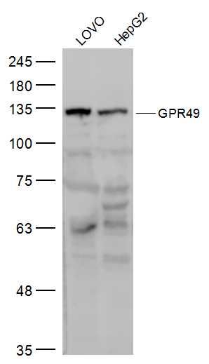 Sample:
LOVO(Human) Cell Lysate at 30 ug
HepG2(Human) Cell Lysate at 30 ug
Primary: Anti-GPR49 (bs-1117R) at 1/500 dilution
Secondary: IRDye800CW Goat Anti-Rabbit IgG at 1/20000 dilution
Predicted band size: 98 kD
Observed band size: 125 kD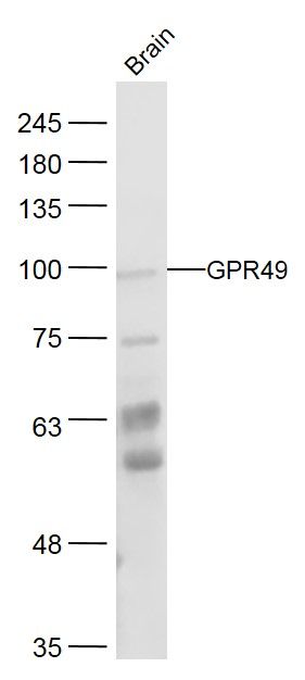 Sample:
Brain (Mouse) Lysate at 40 ug
Primary: Anti-GPR49 (bs-1117R) at 1/300 dilution
Secondary: IRDye800CW Goat Anti-Rabbit IgG at 1/20000 dilution
Predicted band size: 98 kD
Observed band size: 98 kD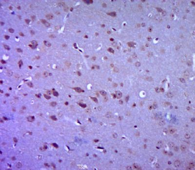 Paraformaldehyde-fixed, paraffin embedded (mouse brain tissue); Antigen retrieval by boiling in sodium citrate buffer (pH6.0) for 15min; Block endogenous peroxidase by 3% hydrogen peroxide for 20 minutes; Blocking buffer (normal goat serum) at 37°C for 30min; Antibody incubation with (GPR49) Polyclonal Antibody, Unconjugated (bs-1117R) at 1:400 overnight at 4°C, followed by a conjugated secondary (sp-0023) for 20 minutes and DAB staining.
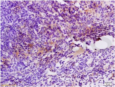 Tissue/cell: mouse colon carcinoma; 4% Paraformaldehyde-fixed and paraffin-embedded;
Antigen retrieval: citrate buffer ( 0.01M, pH 6.0 ), Boiling bathing for 15min; Block endogenous peroxidase by 3% Hydrogen peroxide for 30min; Blocking buffer (normal goat serum,C-0005) at 37℃ for 20 min;
Incubation: Anti-GPR49/LGR5 Polyclonal Antibody, Unconjugated(bs-1117R) 1:200, overnight at 4°C, followed by conjugation to the secondary antibody(SP-0023) and DAB(C-0010) staining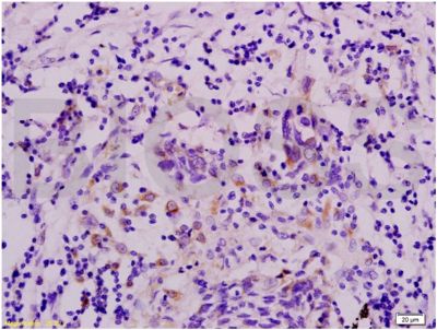 Tissue/cell: human colon carcinoma; 4% Paraformaldehyde-fixed and paraffin-embedded;
Antigen retrieval: citrate buffer ( 0.01M, pH 6.0 ), Boiling bathing for 15min; Block endogenous peroxidase by 3% Hydrogen peroxide for 30min; Blocking buffer (normal goat serum,C-0005) at 37℃ for 20 min;
Incubation: Anti-GPR49/LGR5 Polyclonal Antibody, Unconjugated(bs-1117R) 1:200, overnight at 4°C, followed by conjugation to the secondary antibody(SP-0023) and DAB(C-0010) staining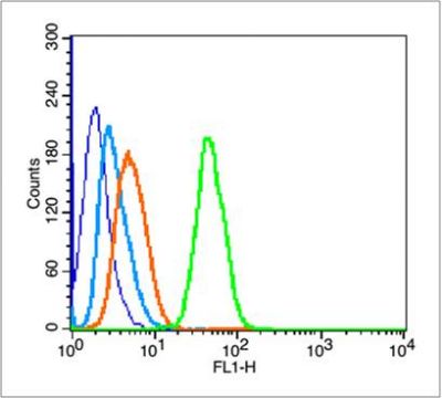 Blank control (blue line):Hela(blue).
Primary Antibody (green line): Rabbit Anti-GPR49 antibody(bs-1117R),
Dilution: 3μg /10^6 cells;
Isotype Control Antibody (orange line): Rabbit IgG .
Secondary Antibody (white blue line): Goat anti-rabbit IgG-FITC
Dilution: 1μg /test.
Protocol
The cells were fixed with 70% ethanol (Overnight at 4℃) and then permeabilized with 90% ice-cold methanol for 30 min on ice. Cells stained with Primary Antibody for 30 min at room temperature. The cells were then incubated in 1 X PBS/2%BSA/10% goat serum to block non-specific protein-protein interactions followed by the antibody for 15 min at room temperature. The secondary antibody used for 40 min at room temperature. Acquisition of 20,000 events was performed.

