产品中心
当前位置:首页>产品中心Anti-FOXO3A
货号: bs-1548R 基本售价: 780.0 元 规格: 50ul
- 规格:50ul
- 价格:780.00元
- 规格:100ul
- 价格:1380.00元
- 规格:200ul
- 价格:2200.00元
产品信息
- 产品编号
- bs-1548R
- 英文名称
- FOXO3A
- 中文名称
- 叉头蛋白O3A抗体
- 别 名
- AF6q21; AF6q21 protein; DKFZp781A0677; FKHR2; FKHRL 1; FKHRL1; FKHRL1P2; Forkhead (Drosophila) homolog (rhabdomyosarcoma) like 1; Forkhead box O3A; Forkhead box protein O3A; Forkhead Drosophila homolog of in rhabdomyosarcoma like 1; Forkhead in rhabdomyosarcoma like 1; FOX O3A; FOXO2; MGC12739; MGC31925; FOXO3_HUMAN

- Specific References (1) | bs-1548R has been referenced in 1 publications.[IF=4.97] Morales, María Gabriela, et al. "Angiotensin-(1-7) attenuates disuse skeletal muscle atrophy via the Mas receptor." Disease Models and Mechanisms(2016): dmm-023390. WB ; Mouse.PubMed:26851244
- 规格价格
- 50ul/780元购买 100ul/1380元购买 200ul/2200元购买 大包装/询价
- 说 明 书
- 50ul 100ul 200ul
- 研究领域
- 肿瘤 免疫学 转录调节因子
- 抗体来源
- Rabbit
- 克隆类型
- Polyclonal
- 交叉反应
- Human, Mouse, Rat, Chicken, Dog, Pig, Cow, Horse, Sheep,
- 产品应用
- WB=1:500-2000 ELISA=1:500-1000 IHC-P=1:400-800 IHC-F=1:400-800 Flow-Cyt=1ug/Test IF=1:100-500 (石蜡切片需做抗原修复)
not yet tested in other applications.
optimal dilutions/concentrations should be determined by the end user.
- 分 子 量
- 74kDa
- 细胞定位
- 细胞核 细胞浆
- 性 状
- Lyophilized or Liquid
- 浓 度
- 1mg/ml
- 免 疫 原
- KLH conjugated synthetic peptide derived from human FOXO3A:581-673/673
- 亚 型
- IgG
- 纯化方法
- affinity purified by Protein A
- 储 存 液
- 0.01M TBS(pH7.4) with 1% BSA, 0.03% Proclin300 and 50% Glycerol.
- 保存条件
- Store at -20 °C for one year. Avoid repeated freeze/thaw cycles. The lyophilized antibody is stable at room temperature for at least one month and for greater than a year when kept at -20°C. When reconstituted in sterile pH 7.4 0.01M PBS or diluent of antibody the antibody is stable for at least two weeks at 2-4 °C.
- PubMed
- PubMed
- 产品介绍
- background:
This gene belongs to the forkhead family of transcription factors which are characterized by a distinct forkhead domain. This gene likely functions as a trigger for apoptosis through expression of genes necessary for cell death. Translocation of this gene with the MLL gene is associated with secondary acute leukemia. Alternatively spliced transcript variants encoding the same protein have been observed. [provided by RefSeq, Jul 2008]
Function:
Transcriptional activator which triggers apoptosis in the absence of survival factors, including neuronal cell death upon oxidative stress. Recognizes and binds to the DNA sequence 5-[AG]TAAA[TC]A-3. Participates in post-transcriptional regulation of MYC: following phosphorylation by MAPKAPK5, promotes induction of miR-34b and miR-34c expression, 2 post-transcriptional regulators of MYC that bind to the 3UTR of MYC transcript and prevent its translation.
Subunit:
Interacts with YWHAB/14-3-3-beta and YWHAZ/14-3-3-zeta, which are required for cytosolic sequestration. Upon oxidative stress, interacts with STK4/MST1, which disrupts interaction with YWHAB/14-3-3-beta and leads to nuclear translocation. Interacts with PIM1.
Subcellular Location:
Cytoplasm, cytosol. Nucleus. Note=Translocates to the nucleus upon oxidative stress and in the absence of survival factors.
Tissue Specificity:
Ubiquitous.
Post-translational modifications:
In the presence of survival factors such as IGF-1, phosphorylated on Thr-32 and Ser-253 by AKT1/PKB. This phosphorylated form then interacts with 14-3-3 proteins and is retained in the cytoplasm. Survival factor withdrawal induces dephosphorylation and promotes translocation to the nucleus where the dephosphorylated protein induces transcription of target genes and triggers apoptosis. Although AKT1/PKB doesnt appear to phosphorylate Ser-315 directly, it may activate other kinases that trigger phosphorylation at this residue. Phosphorylated by STK4/MST1 on Ser-209 upon oxidative stress, which leads to dissociation from YWHAB/14-3-3-beta and nuclear translocation. Phosphorylated by PIM1. Phosphorylation by AMPK leads to the activation of transcriptional activity without affecting subcellular localization. Phosphorylation by MAPKAPK5 promotes nuclear localization and DNA-binding, leading to induction of miR-34b and miR-34c expression, 2 post-transcriptional regulators of MYC that bind to the 3UTR of MYC transcript and prevent its translation.
DISEASE:
Note=A chromosomal aberration involving FOXO3 is found in secondary acute leukemias. Translocation t(6;11)(q21;q23) with MLL/HRX.
Similarity:
Contains 1 fork-head DNA-binding domain.
SWISS:
O43524
Gene ID:
2309
Database links:Entrez Gene: 2309 Human
Entrez Gene: 56484 Mouse
Entrez Gene: 294515 Rat
Omim: 602681 Human
SwissProt: O43524 Human
SwissProt: Q9WVH4 Mouse
Unigene: 220950 Human
Unigene: 338613 Mouse
Unigene: 24593 Rat
Important Note:
This product as supplied is intended for research use only, not for use in human, therapeutic or diagnostic applications.
FOXO3a是FOX(forkhead box)蛋白家族的一个重要成员,与细胞转化、肿瘤的发生发展及其血管生成有关,是一种已知的控制细胞循环和细胞死亡的蛋白质。
- 产品图片
 Sample:
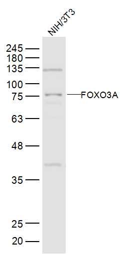
NIH/3T3(Mouse) Cell Lysate at 30 ug
Primary: Anti-FOXO3A (bs-1548R) at 1/500 dilution
Secondary: IRDye800CW Goat Anti-Rabbit IgG at 1/20000 dilution
Predicted band size: 74 kD
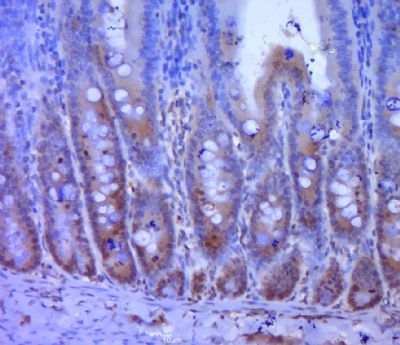
Observed band size: 75 kD Paraformaldehyde-fixed, paraffin embedded (Rat rectum tissue); Antigen retrieval by boiling in sodium citrate buffer (pH6.0) for 15min; Block endogenous peroxidase by 3% hydrogen peroxide for 20 minutes; Blocking buffer (normal goat serum) at 37°C for 30min; Antibody incubation with (FOXO3A) Polyclonal Antibody, Unconjugated (bs-1548R) at 1:400 overnight at 4°C, followed by a conjugated secondary (sp-0023) for 20 minutes and DAB staining.
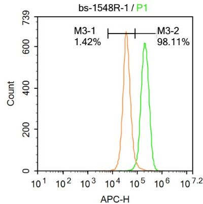
 Blank control: A431.
Primary Antibody (green line): Rabbit Anti-FOXO3A antibody (bs-1548R)
Dilution: 1μg /10^6 cells;
Isotype Control Antibody (orange line): Rabbit IgG .
Secondary Antibody : Goat anti-rabbit IgG-AF647
Dilution: 1μg /test.
Protocol
The cells were fixed with 4% PFA (10min at room temperature)and then permeabilized with 90% ice-cold methanol for 20 min at-20℃. The cells were then incubated in 5%BSA to block non-specific protein-protein interactions for 30 min at at room temperature .Cells stained with Primary Antibody for 30 min at room temperature. The secondary antibody used for 40 min at room temperature. Acquisition of 20,000 events was performed.

