

产品中心
当前位置:首页>产品中心Anti-MAP1LC3A
货号: bs-1534R 基本售价: 780.0 元 规格: 50ul
- 规格:50ul
- 价格:780.00元
- 规格:100ul
- 价格:1380.00元
- 规格:200ul
- 价格:2200.00元
产品信息
- 产品编号
- bs-1534R
- 英文名称
- MAP1LC3A
- 中文名称
- 自噬微管相关蛋白轻链3抗体
- 别 名
- ATG8E; Autophagy-related protein LC3 A; Autophagy-related ubiquitin-like modifier LC3 A; LC3; LC3A; MAP1 light chain 3 like protein 1; MAP1 light chain 3-like protein 1; MAP1A/1B light chain 3 A; MAP1A/MAP1B LC3 A; MAP1A/MAP1B light chain 3 A; MAP1ALC3; MAP1BLC3; Map1lc3a; Microtubule associated proteins 1A/1B light chain 3; Microtubule-associated protein 1 light chain 3 alpha; Microtubule-associated proteins 1A and 1B, light chain 3; Microtubule-associated proteins 1A/1B light chain 3A; MLP3A_HUMAN.
- 规格价格
- 50ul/780元购买 100ul/1380元购买 200ul/2200元购买 大包装/询价
- 说 明 书
- 50ul 100ul 200ul
- 研究领域
- 肿瘤 细胞生物 免疫学 神经生物学 信号转导 细胞凋亡 激酶和磷酸酶 细胞自噬
- 抗体来源
- Rabbit
- 克隆类型
- Polyclonal
- 交叉反应
- Human, Mouse, Rat, Chicken, Pig, Cow,
- 产品应用
- WB=1:500-2000 ELISA=1:500-1000 IHC-P=1:400-800 IHC-F=1:400-800 Flow-Cyt=0.2μg/Test IF=1:100-500 (石蜡切片需做抗原修复)
not yet tested in other applications.
optimal dilutions/concentrations should be determined by the end user.
- 分 子 量
- 14kDa
- 细胞定位
- 细胞浆 细胞膜
- 性 状
- Lyophilized or Liquid
- 浓 度
- 1mg/ml
- 免 疫 原
- KLH conjugated synthetic peptide derived from human MAP1LC3A:75-121/121
- 亚 型
- IgG
- 纯化方法
- affinity purified by Protein A
- 储 存 液
- 0.01M TBS(pH7.4) with 1% BSA, 0.03% Proclin300 and 50% Glycerol.
- 保存条件
- Store at -20 °C for one year. Avoid repeated freeze/thaw cycles. The lyophilized antibody is stable at room temperature for at least one month and for greater than a year when kept at -20°C. When reconstituted in sterile pH 7.4 0.01M PBS or diluent of antibody the antibody is stable for at least two weeks at 2-4 °C.
- PubMed
- PubMed
- 产品介绍
- background:
Microtubule-associated MAPILC3A constitutes nearly half of the mass of all the microtubule associated proteins that copurify with brain microtubules. MAP1LC3A is one of three human orthologs of the rat Map1LC3, (named MAP1LC3A, MAP1LC3B, and MAP1LC3C). The three isoforms of human MAP1LC3 exhibit distinct expression patterns in different human tissues and also differ in their post-translation modifications. MAP1LC3A and MAP1LC3C are produced by the proteolytic cleavage after the conserved C-terminal Gly residue; MAP1LC3B does not undergo C-terminal cleavage and exists in a single modified form.
Function:
Probably involved in formation of autophagosomal vacuoles (autophagosomes).
Subunit:
3 different light chains, LC1, LC2 and LC3, can associate with MAP1A and MAP1B proteins. Interacts with SQSTM1. Interacts with TP53INP1 and TP53INP2.
Subcellular Location:
Cytoplasm, cytoskeleton. Endomembrane system; Lipid-anchor. Cytoplasmic vesicle, autophagosome membrane; Lipid-anchor. Cytoplasmic vesicle, autophagosome. Note=LC3-II binds to the autophagic membranes.
Tissue Specificity:
Most abundant in heart, brain, liver, skeletal muscle and testis but absent in thymus and peripheral blood leukocytes.
Post-translational modifications:
The precursor molecule is cleaved by APG4B/ATG4B to form the cytosolic form, LC3-I. This is activated by APG7L/ATG7, transferred to ATG3 and conjugated to phospholipid to form the membrane-bound form, LC3-II.
Similarity:
Detects a band of approximately 16 kDa (predicted molecular weight: 14 kDa).
SWISS:
Q9H492
Gene ID:
84557
Database links:Entrez Gene: 84557 Human
Entrez Gene: 66734 Mouse
Entrez Gene: 362245 Rat
Omim: 601242 Human
SwissProt: Q9H492 Human
SwissProt: Q91VR7 Mouse
SwissProt: Q6XVN8 Rat
Unigene: 632273 Human
Unigene: 196239 Mouse
Unigene: 3135 Rat
Important Note:
This product as supplied is intended for research use only, not for use in human, therapeutic or diagnostic applications.
- 产品图片
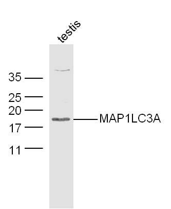 Sample:Testis (Mouse) Lysate at 30 ug
Primary: Anti-MAP1LC3A (bs-1534R) at 1/300 dilution
Secondary: IRDye800CW Goat Anti-Rabbit IgG at 1/20000 dilution
Predicted band size: 14 kD
Observed band size: 18 kD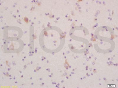 Tissue/cell: human glioma tissue; 4% Paraformaldehyde-fixed and paraffin-embedded;
Antigen retrieval: citrate buffer ( 0.01M, pH 6.0 ), Boiling bathing for 15min; Block endogenous peroxidase by 3% Hydrogen peroxide for 30min; Blocking buffer (normal goat serum,C-0005) at 37℃ for 20 min;
Incubation: Anti-LC3 α/MAP1A/MAP LC3 Alpha/Beta Polyclonal Antibody, Unconjugated (bs-1534R) 1:200, overnight at 4°C, followed by conjugation to the secondary antibody(SP-0023) and DAB(C-0010) staining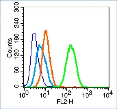 Blank control (blue line): Hela (blue).
Primary Antibody (green line): Rabbit Anti-MAP1LC3A antibody (bs-1534R)
Dilution: 0.2μg /10^6 cells;
Isotype Control Antibody (orange line): Rabbit IgG .
Secondary Antibody (white blue line): Goat anti-rabbit IgG-PE
Dilution: 1μg /test.
Protocol
The cells were fixed with 70% ethanol (Overmight at 4℃) and then permeabilized with 90% ice-cold methanol for 30 min at -20℃.Cells stained with Primary Antibody for 30 min at room temperature. The cells were then incubated in 1 X PBS/2%BSA/10% goat serum to block non-specific protein-protein interactions followed by the antibody for 15 min at room temperature. The secondary antibody used for 40 min at room temperature. Acquisition of 20,000 events was performed.